Adenomas are benign tumors arising from glandular epithelium, or from other non-glandular tissues, if the tumor has a gland-like appearance. In the case of a meibomian adenoma, the origin is in the meibomian glands of the eyelid. The meibomian glands are modified sebaceous glands, serving to produce some of the protective film that covers the eyeball. They're named for Heinrich Meibom (1638-1700), a German anatomist and ophthalmologist. They are also referred to as "tarsal glands."
 As is true of all sebaceous glands and their derivatives, the process of secretion involves the proliferation of cells at the periphery of the gland, and their steady progression towards the center, accompanied by their dissolution to produce the secretory product. The cells are proliferative, a feature of most epithelia. Some epithelia (most clearly exemplified by the skin and the lining of the digestive tract) proliferate at a very high rate, but some, like these glands, are much more moderate and controlled. In the case of sebaceous gland derivatives the rate of proliferation has to match that of secretion.
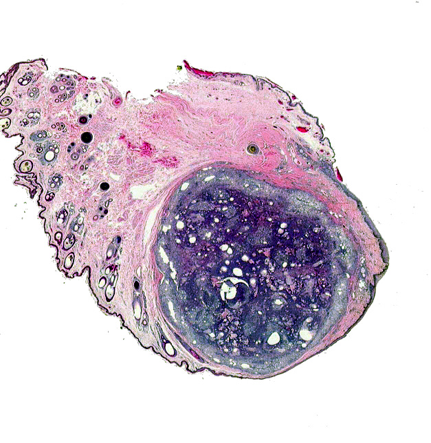
Proliferation, however, in all cases, has to be under complete control. In this example the control has been lost. Something has occurred to trigger proliferation that's far too rapid, and the end result is that the gland(s) increase in size, finally manifesting themselves as visible growths on the exterior of the eyelid. If this continues unabated, there is potential to injure the cornea by abrasion. The cure is to remove the mass surgically before it gets too large. At left is the mass excised from Brutus' eye. The tumor is well-defined and located in between the inner (conjunctival) surface of the eyelid and the outer (dermal) side. Other, unaffected glands and some hair follicles, are also present.
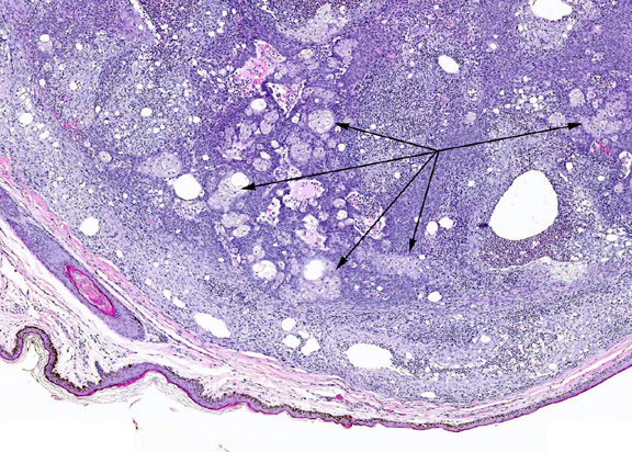
 Another feature of the meibomian gland is secretion; this is, after all, glandular epithelium. Secretion requires the cell to engage in specific activities, hence it has to be differentiated properly. The meibomian gland adenoma's histological appearance after excision is more or less that of an otherwise-normal sebaceous gland derivative. It's got all the proper pathways and activities to do what it's supposed to do, but there's too much of it. At left, you can see several areas of well-differentiated sebaceous-like glandular epithelium, and at high magnification (scroll over) the normal appearance of these cells is pretty obvious. Sebaceous glands are discussed in general terms in Exercise 14.
Brutus has a benign tumor. Because this tumor is well differentiated, it doesn't have the potential to invade surrounding tissues and/or spread through the blood or lymph systems (metastasis). In general, the more differentiated the cells of a tumor are, the less potential it has for spreading. But it will continue to grow, and depending on where it is can do damage. Remember: "Benign" doesn't mean "harmless," it means "non-invasive." Obviously if the tumor gets large enough it can (and will) cause mechanical damage to the eyeball.
References:
McGavin, M.D., and J.F. Zachary. 2007. "Epithelial Tumors " and "Neoplasms of the Eyelids" IN: Pathologic Basis of Veterinary Disease (4th Edition). Mosby/Elsevier (St. Louis). ISBN 13-978-0-323-02870-7. Pp. 57-58 and 1408-1409.
Ishikawa, K., H. Sakai, M. Hosoi, G. Ohta, T. Yanai, and T. Masegi. 2005. Immunohistochemical demonstration of S-phase cells in canine and feline spontaneous tumors by anti-bromodeoxyuridine monoclonal antibody. Journal of Toxicological Pathology 18:135-140.
Pakhrin, B. M-S. Kang, I-H. Bae, M-S. Park, H. Jee, M-H. You, J-H. Kim, B-I. Yoon, Y-K. Choi, and D-Y. Kim. 2007. Retrospective study of canine cutaneous tumors in Korea. Journal of Veterinary Science 8:229-236.
 As is true of all sebaceous glands and their derivatives, the process of secretion involves the proliferation of cells at the periphery of the gland, and their steady progression towards the center, accompanied by their dissolution to produce the secretory product. The cells are proliferative, a feature of most epithelia. Some epithelia (most clearly exemplified by the skin and the lining of the digestive tract) proliferate at a very high rate, but some, like these glands, are much more moderate and controlled. In the case of sebaceous gland derivatives the rate of proliferation has to match that of secretion.