
The retina is the whole point of the eye: everything else is there to support it, provide for its needs, and protect it, so that it can respond to light. The retina is a fantastically complex array of neural and glial elements. It's structurally an extension of the brain, at least in part. Its innermost surface is overlain by the vitreous body; its outermost portion is right up against the uveal tunic.
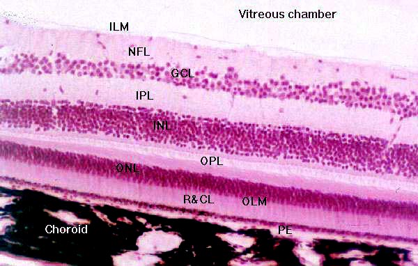
Here is a section through the retina. The different layers are marked for clarity. Remember that the light has to pass through the entire structure before reaching the sensitive elements, so it's coming from the top of the image.
At the top of the screen is the vitreous chamber, and marking off the innermost limit of the retina is the inner limiting membrane (ILM). This isn't a membrane, despite the name. It's the fused feet of the Müller cells, the retina's glial element.
Outwards from there is the nerve fiber layer (NFL), where axons of the ganglion cells are bundled together to form the origins of the optic nerve. These fibers are derived from cell bodies of ganglion cells, located in the ganglion cell layer (GCL). The ganglion cells are the final neuron in the chain that sends information to the visual nucleus.
The ganglion cells take their input via synapses in the inner plexiform layer (IPL). Ganglion cell dendrites join there with axons from bipolar cells. The bipolar cells are neurons whose cell bodies comprise the inner nuclear layer (INL). Integrator neurons (the horizontal and amacrine cells) also have their bodies in the inner nuclear layer.
The outer plexiform layer (OPL) contains the dendrites of the bipolar cells, and their synapses with the axons of rod and cone cells. The somata of the rods and cones reside in the outer nuclear layer (ONL).
The actual light-sensitive parts of the rods and cones are separated from the rest of the system. by the outer limiting membrane (OLM). Again, this isn't a membrane: it's a region of occluding junctions, this time between Müller cells and the the rod and cone cells.
Finally, the layer of rods and cones (R&CL) are the actual light sensitive elements. The pigment
epithelium layer (PE) is strictly speaking not part of the
retina proper, but some texts will include it as such. It's roles include phagocytosis of the used-up rod and cone material (which is replaced) and participation in the cycle by which visual pigments are formed. The retinal pigment layer forms part of the blood-retina barrier and is a major regulator of immune function in the outer retina.