Comparison of Salivary Gland Structures
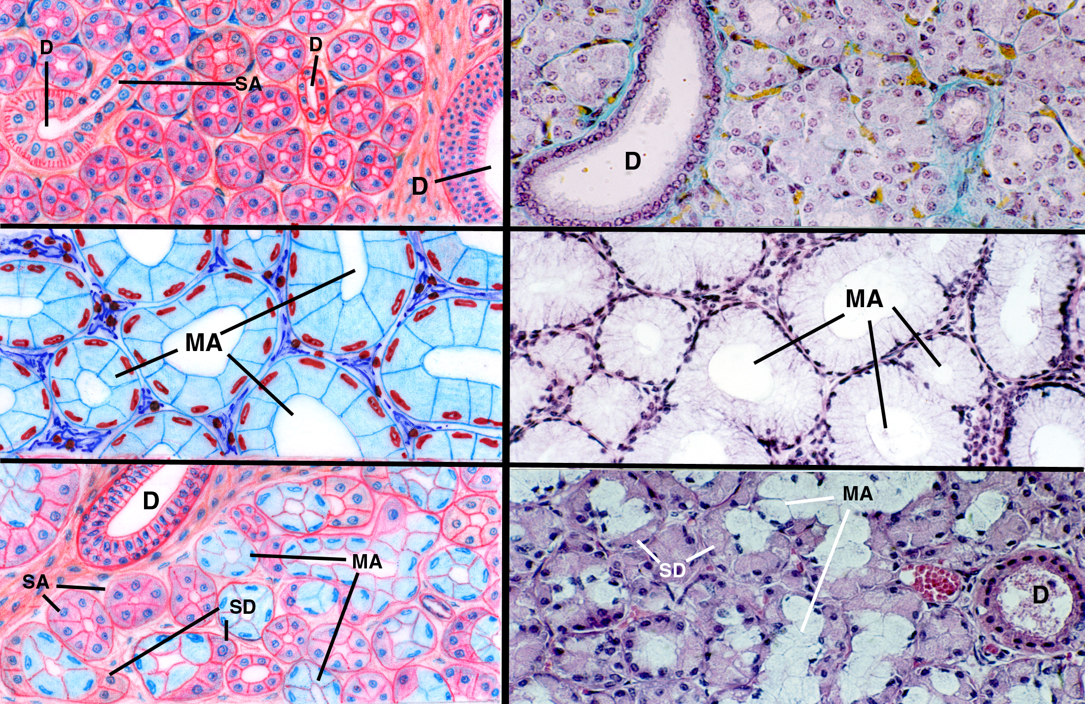
This composite image is worth examining carefully, as it shows the principal arrangements of salivary glands as found in most animals. The three images on the left are idealized drawings; and those on the right are actual micrographs taken from real specimens that correspond to the types in the sketch.
All salivary glands are structurally compound exocrine glands; that is, they have a duct system that takes secretions from the productive areas to wherever they're destined to be used. The duct system is divided. A section through such a gland will almost always show a duct of some kind, as do the images above. Some ducts are fairly small, some are much larger. Regardless of the type of gland and the secretion it makes, s the normal pattern for ductwork is that small ones are lined by simple cuboidal epithelium (above, right, about 500x), and the largest ones are lined by stratified epithelium which may be cuboidal or columnar in shape (above, left, about 100x). The duct system is like a watershed. Small streams join with other small streams to form larger ones, which in turn join to form rivers, until eventually a very large river drains the entire area. The smallest ducts drain one secretory area, and the larger ones take input from more and more of the gland until finally the single main duct takes all of the output. The secretory regions are the most "upstream" ends of the system and to continue the watershed analogy, they are the "springs" from which the streams take their source.
 It is also clear from casual observation that
the secretory regions differ in appearance. This is due to the type of secretions
they make and the viscosity of the secretions. The two top illustrations are
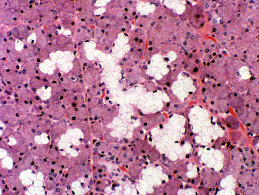
of a serous gland: the secretions of this type of gland are watery
and of low viscosity. The cells of the serous acini (SA) tend to stain
fairly heavily with H&E, and to have a wedge-shaped appearance. Note that
the nuclei of serous acinar cells is round and centrally located in the cytoplasm.
Compare these cells to the ones in the middle set of images: these are mucous
acini (MA). In glands producing a mucous secretion the product is quite
viscous, and it tends to flatten the nuclei out and push them to the periphery
of the acinus. It also isn't very responsive to an H&E stain, but it's
strongly PAS-positive.
It is also clear from casual observation that
the secretory regions differ in appearance. This is due to the type of secretions
they make and the viscosity of the secretions. The two top illustrations are
of a serous gland: the secretions of this type of gland are watery
and of low viscosity. The cells of the serous acini (SA) tend to stain
fairly heavily with H&E, and to have a wedge-shaped appearance. Note that
the nuclei of serous acinar cells is round and centrally located in the cytoplasm.
Compare these cells to the ones in the middle set of images: these are mucous
acini (MA). In glands producing a mucous secretion the product is quite
viscous, and it tends to flatten the nuclei out and push them to the periphery
of the acinus. It also isn't very responsive to an H&E stain, but it's
strongly PAS-positive.
Some salivary glands produce both mucous and serous secretions, and these are called "mixed" glands, for obvious reasons. The bottom images in the composite above reflect this situation, and they show the possible arrangements by which the two secretory areas can be combined. In this sort of mixed gland, the mucous acini are usually pretty obvious. The serous acini can be entirely separated from the mucous acini, but they may also form a sort of "cap" over the end of a mucous acinus. This "cap" is a serous demilune, and in this situation the secretions are mixed together right from the start.
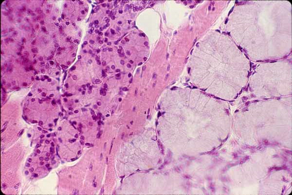
Another example of a mixed gland, this one in which the serous and mucous segments are clearly separated, is shown at left. This is a fairly common situation; in fact, in domestic animals, nearly all salivary glands are mixed to some degree: with the possible exception of the parotid gland (alleged in some references to be purely serous) it's doubtful if you could find a gland that didn't have some elements of both types in it. Whatever the structural arrangements of the gland, the ductwork is the same for both types of secretion.


Some salivary glands are embedded in the wall of the oral cavity, and some are embedded in the tongue. these latter are called lingual salivary glands because of their location. Don't confuse them with the "sublingual salivary gland," a discrete organ in and of itself. Lingual glands may be of the serous or mucous or mixed type, and are scattered through the substance of the tongue. Some of them open out via ducts onto the surface, some of them have ductwork leading to the "moat" that surrounds the large tongue papillae. The images above illustrate this. In the left panel you see both serous and mucous secretory areas, in among the strands of skeletal muscle in the tongue. At right, a large serous gland is discharging via a duct (D) into the moat (M) around a vallate papilla.
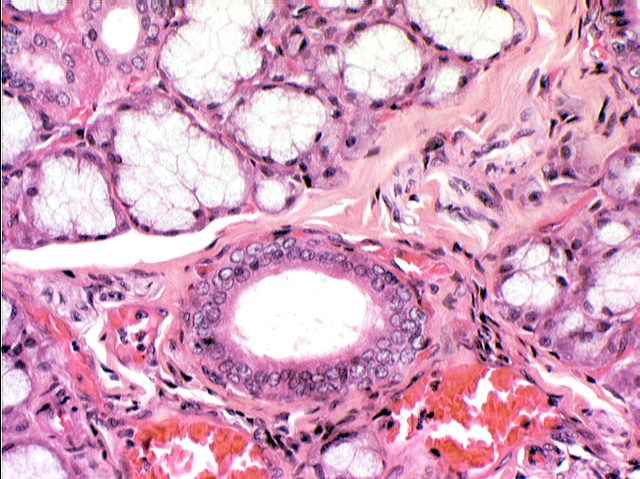
Salivary glands may also be described by the shape of the secretory regions. Most of them are referred to as acinar or tubuloacinar, meaning that the secretory areas are round to oval shaped (acinar) or somewhat elongated, with bulbous ends (tubuloacinar). An example is shown below. At left you see the gland at low magnification: in this image the numerous duct profiles of this gland are evident. Note that this must be a compound gland. Why? Because, since a gross dissection of the organ would show that there is only one main duct carrying all the secretions, all of these smaller duct profiles must eventually lead into it. hence, the duct system is divided, and therefore compound.

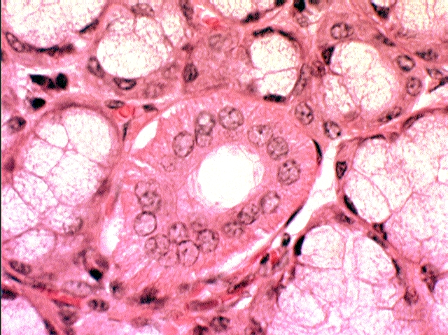
At right, at somewhat higher magnification, a secretory region of the tubuloacinar shape is highlighted. The "tubulo-" section is the relatively straight part, and the bulbous termini are the "acinar" parts. For comparison, some of the acinar secretory regions of this gland (which are producing a serous output) are marked as well. Remember these structures have depth. The acinar parts are shaped like grapes or eggs; the tubuloacinar parts are shaped like...well, maybe a knobby banana would be the best comparison.
So, to sum up: the complete description of this gland would be:
COMPOUND, TUBULOACINAR, MIXED SEROUS & MUCOUS
One final point: some books will tell you there's "no such thing as a serous tubuloacinar gland." That's not correct. They're few and far between, but there's at least one example, the lachrymal gland of the eye.
*That's a plural. One acinus, two acini. The word comes from the Latin word for a berry or grape. The shape of these things is roughly that of a grape; hence when cut in cross section they appear round, in longitudinal section somewhat elongated.