VM8054 Veterinary Histology
Example: Fundic Glands
Author: Dr. Thomas Caceci


These images are vertical sections through the glandular portion
of the fundic stomach. On the left you see a low-power view through a number
of glands; the parietal cells (PC) are easily seen as large, eosinophilic
cells scattered in a background of more basophilic chief cells. The fundic
glands are by far the most numerous ones in mammalian stomachs. There are
several million in most species. Like all stomach glands, they're located
in the deep parts of the tunica mucosa, surrounded by the connective tissue
of the lamina propria. The gastric pits, into which the glands discharge their
secretions, are not visible: they've been cropped out of the image at the
top of the field.
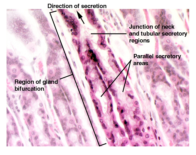
What isn't evident in this image is the actual structure of
the glands themselves, which are of the simple branched tubular shape.
The image on the right has been enhanced to make this more plain. The simple
branched tubular gland is an inverted Y-shape: the "neck" of the
gland is the stem of the Y and it is directed towards the top of the field
(i.e., the free surface and the gastric pit). At the point indicated
by the arrows, the gland bifurcates into two long, tube-shaped secretory
regions, in which reside the chief and parietal cells. These continue to
the deepest levels of the lamina propria and butt up against the muscularis
mucosae. They do NOT extend into the submucosal layer.
The fundic glands are the source of the proteases of the stomach
(trypsin and chymotrypsin) and the hydrochloric acid that gives the stomach
contents its pH of about 2.0. In mammals these are produced by two different
cell types: the parietal cells make the acid, and the chief cells
make the proteases. In other groups of animals (e.g., birds) one cell
type has both functions.
In this image the darkly stained cells are the chief cells (CC).
Chunky and angular in shape, in the electron microscope they show the
typical appearance of protein secreting cells. The parietal
cells (PC) are eosinophilic in their staining response. The difference in staining is related to their functions. The chief cells make proteins, hence they show the typical basophilic properties of cells that do this. The parietal cells are essentially ion pumps. They have no protein secretion function and hence little RNA in their cytoplasm.
Dog stomach; H&E stain, paraffin section, 200x
 This drawing should make clear the relationships between the parietal and chief
cells and the extent to whcih the glands descend into the stomach wall. The
muscularis mucosae marks the limit of the tunica mucosae and its underlying
lamina propria; outside of that the submucosal layer begins. Hence, sinec all
of the gland profiles are on the luminal side of the muscularis mucosae, these
tubular secretory regions are supported and separated by the CT of the lamina
propria. There isn't much of it between the tubes, as the fundic glands are
very numerous and tightly packed; but it's there.
At this depth the tubular nature of the gland may not be evident.
That's because the 3-dimensional glands, cut in 2 dimensions, are passing in
and out of the plane of the section. Hence, if there's a bend, they will appear
to be "round" in a 2-D image. Others, whose longitudinal axis is closer
to the plane of the section, will appear elongated, perhaps oval. But in fact
they're all tubes, cut at different angles.
Drawing by Dr. Samir El-Shafey
 The
architecture of the parietal and chief cells is much easier to see in this image.
The parietal cell (PC) is large, round, and eosinophilic, thanks to its
cytoplasmic complement of mitochondria. Since these cells have a high demand
for ATP to drive the pumps that push sodium out of the cytoplasm into the lumen
of the stomach, mitochondria are abundant.
The
architecture of the parietal and chief cells is much easier to see in this image.
The parietal cell (PC) is large, round, and eosinophilic, thanks to its
cytoplasmic complement of mitochondria. Since these cells have a high demand
for ATP to drive the pumps that push sodium out of the cytoplasm into the lumen
of the stomach, mitochondria are abundant.
The appearance of the parietal cell in the light microscope gives no hint of its extensive network of small surface invaginations. These intracellular canaliculi are blind-ended tubules that enormously increase the surface area of the cell on its luminal surface; sort of like reverse microvilli. Each such channel is a site into which chloride and hyrogen ions are pumped by active transport. From the channels they diffuse into the stomach lumen along a local concentration gradient.
The chief cell (CC) has deeply basophilic basal regions, indicative of the presence of large amounts of RER. At its apical end, the chief cell has granular inclusions; these are the packets of enzymes waiting to be released.

Lab Exercise List


 This drawing should make clear the relationships between the parietal and chief
cells and the extent to whcih the glands descend into the stomach wall. The
muscularis mucosae marks the limit of the tunica mucosae and its underlying
lamina propria; outside of that the submucosal layer begins. Hence, sinec all
of the gland profiles are on the luminal side of the muscularis mucosae, these
tubular secretory regions are supported and separated by the CT of the lamina
propria. There isn't much of it between the tubes, as the fundic glands are
very numerous and tightly packed; but it's there.