MAST CELL TUMOR


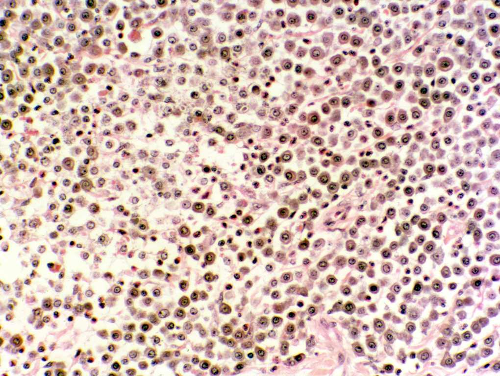
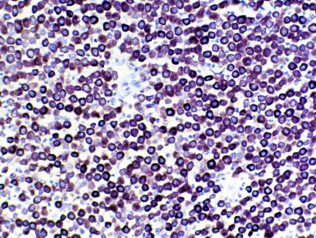
This pair of images emphasizes pretty well how useful a specific stain can be. The image at left is stained with H&E, that at right with TB. Both are of the same specimen: a mast cell tumor from a dog. In fact, they're sections cut from the same block, and photographed at identical magnification (about 200x). The mast cells are far more obvious in the right hand image. They can be easily distinguished from other cell types (e.g., macrophages) by the presence of TB-positive granules. The subtleties and vagaries of the H&E stain reaction make it hard for an inexperienced pathologist to spot mast cells against the "background," but with TB they stand out like neon signs.
Click "Back" to Return to the Example of TB staining