VM8054 Veterinary Histology
Example: Pancreas
Author: Dr. Thomas Caceci
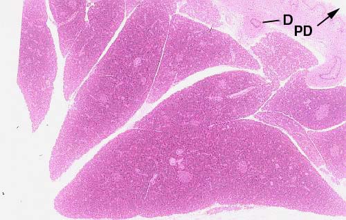
 At low magnification, the pancreas can be seen to be extensively
lobulated. As with other lobular organs, there's a
capsule that surrounds the pancreas; CT septae subdivide
it into lobes and lobules. Each of these eosiniophilic regions is a lobe. Blood
vessels and ducts run through the septae.
The pancreas is a compound acinar gland. An excurrent duct (D)
is visible at the upper right, and this duct is leading into what must be the main pancreatic duct (PD).
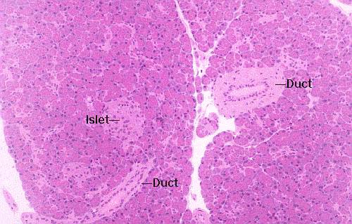
 At
higher magnification the larger ducts in the system are easily visible. They're
lined with a simple columnar epithelium, but this will stratify as the ducts
get large enough.
The output of the exocrine pancreas is channeled through a series of ducts
of increasing caliber, just as in any other exocrine gland, and the nomenclature
is the same. The smallest are the intercalated ducts, each of which serves
only a single secretory acinus. These drain into intralobular ducts,
which in turn drain into interlobular ducts, etc., etc. In most animals
the entire output of digestive enzymes to the intestine is via a single large
pancreatic duct, but an accessory duct is present in some species.
 The
concept of the "acinus" is worth showing in a diagram. The acinus
is the basic secretory unit of the pancreas (and many other exocrine glands,
by the way) and it's composed of secretory cells grouped around a lumen into
which their secretions are released. The word "acinus" is from the
Latin for a "berry" or "grape," and what you're seeing in
this sketch is two acini cut along their long axes.
Each of them is built of the secretory cells that produce the
pancreatic enzymes. These cells show basal basophilia, the deep staining
with hematoxylin that's characteristic of any region with a heavy concentration
of RER. The secretory product of these cells is stored in vesicles derived from
the Golgi apparatus and held in the apical cytoplasm until a signal is received
to release them. At that time they're brought to the surface of the cell, the
granules' membranes fuse with the plasma membrane, and the contents are released
intot eh ductwork by exocytosis. The small duct you see in this sketch will
almost immediately join a larger one.
Note also the so-called "centroacinar cells." These
are nothing more than the very first cells in the duct system. They have no
secretory function. They're the actual beginning of the drainage from a given
acinus.
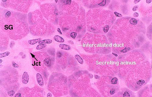
 This
is the most "upstream" end of the system. Here you see a secretory
acinus and its individual intercalated duct, cut longitudinally.
The flow is from right to left in this image. Secretory granules (SG)
are held in the cytoplasm of the acinar cells until the signal to release comes.
The release is by exocytosis and the product begins to flow down the ductwork.
This intercalated duct joins with another at a junction point (Jct) to
form a slightly larger intralobular duct, draining two or more acini.
This
is the most "upstream" end of the system. Here you see a secretory
acinus and its individual intercalated duct, cut longitudinally.
The flow is from right to left in this image. Secretory granules (SG)
are held in the cytoplasm of the acinar cells until the signal to release comes.
The release is by exocytosis and the product begins to flow down the ductwork.
This intercalated duct joins with another at a junction point (Jct) to
form a slightly larger intralobular duct, draining two or more acini.
The secretory product is released into the ducts in an inactive form and become
activated when it reaches the intestine. The reason for this is obvious: the
enzymes in it would destroy the pancreas itself before ever reaching the intestine
if released in the active form.
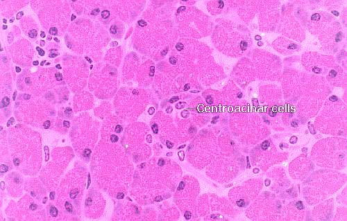
 Frequently
an acinus is cut in such a way as to reveal only the first few cells in the
duct system. These are tucked right up a short way inside the acinus,
rather like the stem on a grape. Since they aren't secretory, they'll be stained
differently than the secretory cells. The classical histology term for these
is the centroacinar cells. They're just the first cells of the intercalated
duct.
Drawing by Dr. Samir El-Shafey
Monkey pancreas; H&E stain, 1.5 µm plastic sections, 20x, 40x,
1000x, and 1000x

Lab Exercise List
 At low magnification, the pancreas can be seen to be extensively
lobulated. As with other lobular organs, there's a
capsule that surrounds the pancreas; CT septae subdivide
it into lobes and lobules. Each of these eosiniophilic regions is a lobe. Blood
vessels and ducts run through the septae.
 At
higher magnification the larger ducts in the system are easily visible. They're
lined with a simple columnar epithelium, but this will stratify as the ducts
get large enough.
  The
concept of the "acinus" is worth showing in a diagram. The acinus
is the basic secretory unit of the pancreas (and many other exocrine glands,
by the way) and it's composed of secretory cells grouped around a lumen into
which their secretions are released. The word "acinus" is from the
Latin for a "berry" or "grape," and what you're seeing in
this sketch is two acini cut along their long axes.
 This
is the most "upstream" end of the system. Here you see a secretory
acinus and its individual intercalated duct, cut longitudinally.
The flow is from right to left in this image. Secretory granules (SG)
are held in the cytoplasm of the acinar cells until the signal to release comes.
The release is by exocytosis and the product begins to flow down the ductwork.
This intercalated duct joins with another at a junction point (Jct) to
form a slightly larger intralobular duct, draining two or more acini.
This
is the most "upstream" end of the system. Here you see a secretory
acinus and its individual intercalated duct, cut longitudinally.
The flow is from right to left in this image. Secretory granules (SG)
are held in the cytoplasm of the acinar cells until the signal to release comes.
The release is by exocytosis and the product begins to flow down the ductwork.
This intercalated duct joins with another at a junction point (Jct) to
form a slightly larger intralobular duct, draining two or more acini.  Frequently
an acinus is cut in such a way as to reveal only the first few cells in the
duct system. These are tucked right up a short way inside the acinus,
rather like the stem on a grape. Since they aren't secretory, they'll be stained
differently than the secretory cells. The classical histology term for these
is the centroacinar cells. They're just the first cells of the intercalated
duct.