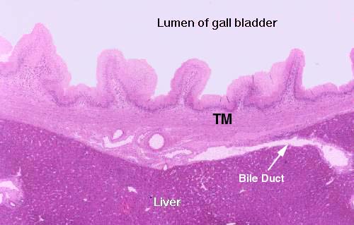
 This low magnification image shows the wall of the gall bladder
pretty well.
What might are first be taken as vill really aren't. They're folds in the mucosa, permanent rugations like those in the stomach. They vary in height depending on how full the bladder is. When it's distended the folds are lower than you see them here.
The gall bladder is only partially covered by a fold of peritoneum; its opposite side is seated in a pocket in the surface of the liver, attached to it by CT. Bile ducts carry bile to the gall bladder for temporary storage.
The tunica muscularis (TM) is rather disorganized, and there is no muscularis mucosae.
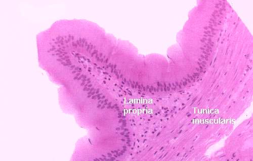
 At higher magnification
you can see the nature of the gall bladder epithelium: a rather uninteresting
simple columnar form, containing no goblet cells. Though these cells do have a
few microvilli on their free surfaces, there is no real brush border.
There isn't any muscularis mucosae, and the tunica muscularis is stringy and scanty and shot through with collagen fibers. Since it has only the function of squirting bile into the duct that carries it to the duodenum, it needn't be well-developed.
Monkey gall bladder; H&E stain, 1.5 µm plastic section, 40x and 200x
