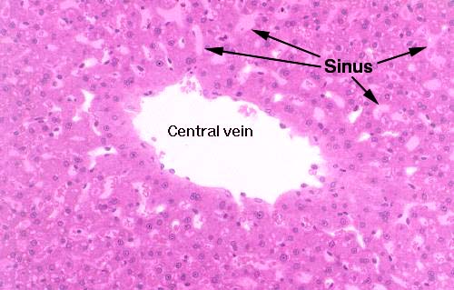
 The hepatocytes of the liver lobule are arrayed in long rows, sort
of like brick walls in which the hepatocytes are the "bricks." The
spaces between the "walls" are the hepatic sinusoids.
Sinusoids are a form of capillary, and it's into these that the blood
is emptied from the arterial and venous input to the liver lobule.
You can see erythrocytes in the spaces between the plates of
hepatocytes and in the central vein as well.
All the sinusoids of a lobule converge on its central vein. The
sinusoids and the central vein are lined with
endothelium (they're blood spaces, after all) and in
this image you can see the endothelial cells in the central vein
quite easily. The lining is discontinuous, and there are places where
hepatocytes are "naked" and exposed directly to the flow of blood
past them. This is important to liver function as it gives the
hepatocytes access to the blood for modification of its constitution.
The discontinuity is much greater in the sinusoids—where the real
action of the liver goes on—than it is in the veins.
