VM8054 Veterinary Histology
Example: Kupffer Cells
Author: Dr. Thomas Caceci
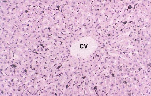 This low power view shows a liver lobule in a rabbit that's been
injected with India ink. The carbon particles of which the ink is
composed have been phagocytosed and the Kupffer cells stand out in
stark contrast to the hepatocytes around them. The radiating
arrangement of the plates of hepatocytes, and the sinusoids between
them, are particularly well demonstrated in this slide. (The unusual
clarity of the architecture in this preparation is probably due to
its having been perfused with fixative; there are no erythrocytes in
the sinusoids because they've been "washed out" by pumping fixative
in via the hepatic artery and portal vein.)
This low power view shows a liver lobule in a rabbit that's been
injected with India ink. The carbon particles of which the ink is
composed have been phagocytosed and the Kupffer cells stand out in
stark contrast to the hepatocytes around them. The radiating
arrangement of the plates of hepatocytes, and the sinusoids between
them, are particularly well demonstrated in this slide. (The unusual
clarity of the architecture in this preparation is probably due to
its having been perfused with fixative; there are no erythrocytes in
the sinusoids because they've been "washed out" by pumping fixative
in via the hepatic artery and portal vein.)
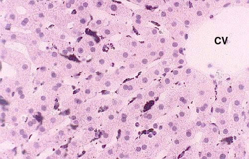 At higher magnification individual Kupffer cells can be made out
pretty well. Note also the relationship of the sinusoids to the
central vein, and the fairly large number of binucleated hepatocytes
that are present in the plates. The image you see here could easily
have come from Metchnikoff's own work, and it's a duplication of one
of the historic experiments in the history of the life sciences,
which proved the reality of phagocytosis.
At higher magnification individual Kupffer cells can be made out
pretty well. Note also the relationship of the sinusoids to the
central vein, and the fairly large number of binucleated hepatocytes
that are present in the plates. The image you see here could easily
have come from Metchnikoff's own work, and it's a duplication of one
of the historic experiments in the history of the life sciences,
which proved the reality of phagocytosis.
Rabbit liver; India ink/Neutral Red stain, paraffin section, 200x and 400x
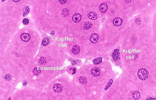 In
this image you can see Kupffer cells in situ, sitting on "top"
of the hepatocytes and actually projecting into the stream of blood oozing past.
The principal role of the Kupffer cell is to remove from the blood any particulate
contaminants that happen to be present, and also to destroy aged erythrocytes.
Their position enables them to do this easily; they just sit there and scarf
up things that come by. These cells actually show some brownish material (probably
lipofuscin) inside them. An eosinophil is also visible. Since the sinusoids
are blood vessels, you can potentially find any of the formed elements of circulating
blood inside them.
In
this image you can see Kupffer cells in situ, sitting on "top"
of the hepatocytes and actually projecting into the stream of blood oozing past.
The principal role of the Kupffer cell is to remove from the blood any particulate
contaminants that happen to be present, and also to destroy aged erythrocytes.
Their position enables them to do this easily; they just sit there and scarf
up things that come by. These cells actually show some brownish material (probably
lipofuscin) inside them. An eosinophil is also visible. Since the sinusoids
are blood vessels, you can potentially find any of the formed elements of circulating
blood inside them.
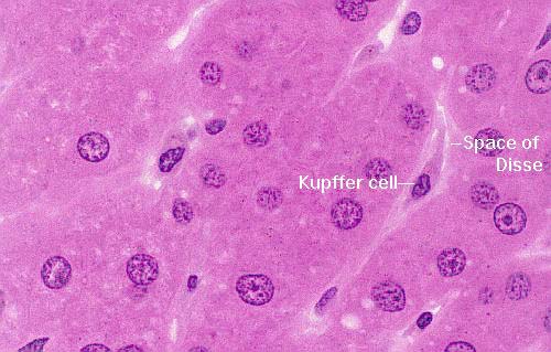 The
Kupffer cell is physically located in the sinusoid, but it's attached
to the hepatocytes, with a small gap between it and the underlying hepatocyte.
This is the Space of Disse (Josef Disse, 1852-1912, a German anatomist).
The space of Disse was a subject of considerable controversy before about 1950
as many people regarded it as an artifact of preparation and believed that the
Kupffer cells (or, alternatively, the endothelial cells if there wasn't a Kupffer
cell around) weren't separated from the hepatocytes. The electron microscope
settled that question: the space is real. Transmission electron micrographs
even show short microvilli on the surface of the hepatocyte that project into
the space of Disse.
The
Kupffer cell is physically located in the sinusoid, but it's attached
to the hepatocytes, with a small gap between it and the underlying hepatocyte.
This is the Space of Disse (Josef Disse, 1852-1912, a German anatomist).
The space of Disse was a subject of considerable controversy before about 1950
as many people regarded it as an artifact of preparation and believed that the
Kupffer cells (or, alternatively, the endothelial cells if there wasn't a Kupffer
cell around) weren't separated from the hepatocytes. The electron microscope
settled that question: the space is real. Transmission electron micrographs
even show short microvilli on the surface of the hepatocyte that project into
the space of Disse.
Monkey liver; H&E stain, 1.5 µm plastic section, 1000x and 1000x

Lab Exercise List