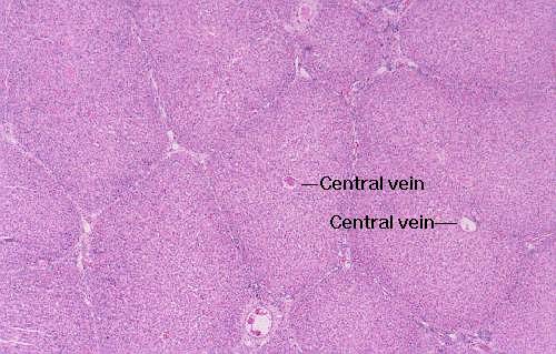
 In this low magnification image the details of the liver's
cellular structure aren't easily seen, but the general outline of the
lobules can be made out without difficulty. This example is from a
pig.
The lobules are roughly hexagonal in shape, and clearly demarcated by CT. In other species the CT content isn't so high as in pigs, and it's a bit harder to make out where one lobule ends and the next begins (see below).
Remember that the lobule is a three dimensional structure, and it projects down into the plane of section, and up out of it, too.
Each lobule has a central vein, the site of drainage of
blood. Blood flows in from the periphery and leaves via the central
vein. Central veins from adjacent lobules coalesce to form larger
vessels, and eventually all of the drainage leaves the liver via the
hepatic vein.
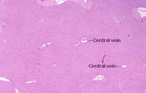
 Here's an example of non-pig liver. The lobulation pattern is much less
distinct the lobulation pattern is compared to that of the pig.
Actually, at moderate magnifications you can pretty easily make out
the boundaries of the lobules by observing the direction of radiation
of the plates of hepatocytes, but the minuscule amount of CT makes it
hard to see the lobulation in a low-power image like this one.
Dog liver; H&E stain, paraffin section, 20x
