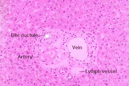
 Here's a classic hepatic triad. These are located at the corners
of the lobules, running through the CT that separates one lobule from
the adjacent ones. These are sites at which blood enters the lobule
and bile leaves it.
Blood comes to the lobule from two sources: the portal vein, a branch of which is shown here as one element of the triad, and the hepatic artery, also shown. The venous input is the larger of the two by a substantial margin; that blood is coming to the liver from the capillary beds in the gut. Hence, it's deoxygenated and nutrient rich. The liver is the first place it goes, and therefore the liver has a chance to detoxify any deleterious materials in the blood before releasing it to the general circulation. The arterial supply carries oxygenated blood, and so in the sinusoidal spaces of the liver lobule, oxygenated and deoxygenated blood are mixed together.
The remaining component of the classical triad is the bile ductule. The one labeled here is a nice example, showing how the wall is composed of simple cuboidal epithelium. The bile ductules carry the exocrine secretion of the liver, bile, that's produced in the hepatocytes and carried to the lumen of the intestine to aid in fat breakdown. A bile ductule like this one will join with others to form larger and larger vessels, which eventually coalesce into one (or, in some species, two) large bile ducts. The flow of bile is outward from the lobule; the flow of blood is inward into it.
In one sense this might perhaps be called a "tetrad" rather than a
triad, because also frequently seen at this point are lymph vessels.
There's one in this image. Lymph vessels drain off the liquid blood
plasma that's released into the intercellular space by flowing blood.
They look like very thin walled veins, but—as you can see
here—unlike veins they never contain erythrocytes under ordinary
circumstances.
