VM8054 Veterinary Histology
Reticulum
Author: Dr. Thomas Caceci
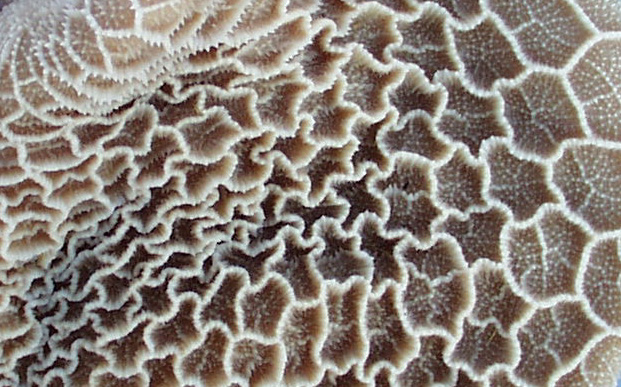 This
scanning EM image makes the "waffle" nature of the reticulum's mucosa
plainer than any words could. Each of the spaces you see here is an epithelium-lined
"pocket." The walls of each pocket contain the muscularis mucosa of
this organ in their tips only. The muscularis mucosae runs around the top edge,
in the CT underlying the epithelium, rather like a drawstring on a purse. When
it contracts, the opening into each pocket is made smaller.
This
scanning EM image makes the "waffle" nature of the reticulum's mucosa
plainer than any words could. Each of the spaces you see here is an epithelium-lined
"pocket." The walls of each pocket contain the muscularis mucosa of
this organ in their tips only. The muscularis mucosae runs around the top edge,
in the CT underlying the epithelium, rather like a drawstring on a purse. When
it contracts, the opening into each pocket is made smaller.
Picture credit: I am indebted to Dr. Mohammed Khalil of
Purdue University's College of Veterinary Medicine for this image.
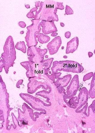 This
view of the reticulum hardly does it justice, but if you relate what it shows
to the SEM image above, you'll see that there are main, or primary folds, arising
from the wall; and that these give rise to secondary folds. In a two-dimensional
histological section like this one, the "waffle" construction isn't
too obvious. If you were standing inside it looking at the wall en face
you'd see pockets lined with epithelium. What you see here is one of those pockets,
cut vertically.
This
view of the reticulum hardly does it justice, but if you relate what it shows
to the SEM image above, you'll see that there are main, or primary folds, arising
from the wall; and that these give rise to secondary folds. In a two-dimensional
histological section like this one, the "waffle" construction isn't
too obvious. If you were standing inside it looking at the wall en face
you'd see pockets lined with epithelium. What you see here is one of those pockets,
cut vertically.
The muscularis mucosae runs along the top edge of the walls of each "pocket"
and connects it to the adjacent ones. Its contractions are thought to help in
moving material in and out of the pockets to facilitate mixing and churning
and hence absorption. Somewhat higher magnification (below, left) shows the
"island" of muscularis mucosae in the tips of the primary fold. This
is made even more obvious in the illustrations below.
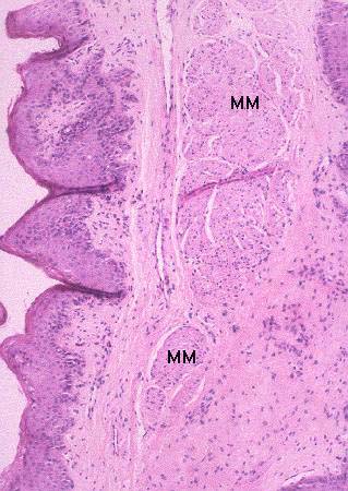
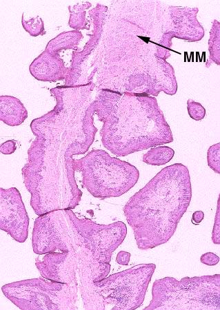
Bovine reticulum; H&E stain, paraffin sections, 20x, 40x, and 100x

Close This Window