VM8054 Veterinary Histology
Omasum
Author: Dr. Thomas Caceci
 The
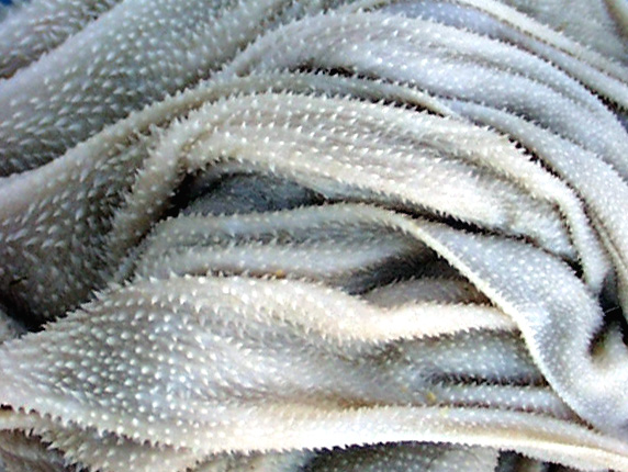
omasum is sometimes called the "butcher's bible" because its mucosa
is formed into leaf-like folds, or foliae, somewhat resembling the leaves of a
book. In histological sections like this one, they appear similar to the papillae
of the rumen, but they're actually flattened in three dimensional view.
The
omasum is sometimes called the "butcher's bible" because its mucosa
is formed into leaf-like folds, or foliae, somewhat resembling the leaves of a
book. In histological sections like this one, they appear similar to the papillae
of the rumen, but they're actually flattened in three dimensional view.
Picture Credit: I am indebted to Dr. Mohammed Khalil of
Purdue University's College of Veterinary Medicine for this striking scanning
EM photo.
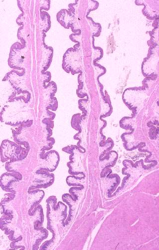 The
omasum's mucosal epithelium, as is the case in other regions of the forestomach,
is a keratinized stratified squamous type. The omasum, however, unlike the rumen
and reticulum, has a true muscularis mucosae. As you can see here, there's a
distinct "core" of muscle in each of the folds. Only part of this
is muscularis mucosae, however; some of it is the tunica muscularis.
The
omasum's mucosal epithelium, as is the case in other regions of the forestomach,
is a keratinized stratified squamous type. The omasum, however, unlike the rumen
and reticulum, has a true muscularis mucosae. As you can see here, there's a
distinct "core" of muscle in each of the folds. Only part of this
is muscularis mucosae, however; some of it is the tunica muscularis.
Even in this low-magnification image you can see that there are three distinct
bands of smooth muscle in the core of each fold. The central one is not muscularis
mucosae. It's part of the tunica muscularis. This really is a flat sheet of
smooth muscle extending up all the way to the tip of the omasal fold, and it's
sandwiched between two parts of the true muscularis mucosae. It's easier to
see this in the high-magnification image below.
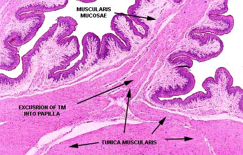
In
this image it becomes more obvious that the innermost band of muscle
fibers in each of these folds is really part of the tunica muscularis. Specifically,
the inner layer of the tunica muscularis sends strands of smooth muscle up into
the fold. These fibers are separated from the muscularis mucosae by a very scanty—but
nevertheless present—submucosa.
Bovine omasum; H&E stain, paraffin sections, 20x and 40x

Close This Window