VM8054 Veterinary Histology
Rumen Papillae
Author: Dr. Thomas Caceci
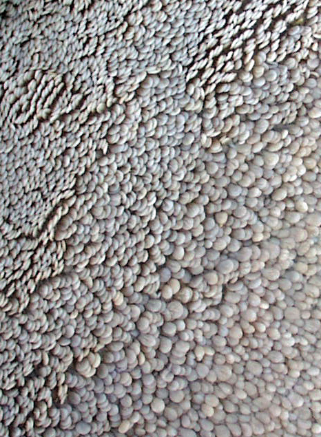 Here
are two views of the rumen papillae. The one at the left is a scanning
electron micrograph, and the three-dimensionality of this method of visualization
shows the papillae to be long, slender finger-like projections. The conventional
image below demonstrates that they're projections of the tunica mucosa only.
They have a core of CT (the lamina propria) and, as is true of all the divisions
of the ruminant forestomach, are covered with stratified squamous epithelium.
There is no muscularis mucosae in the rumen.
Here
are two views of the rumen papillae. The one at the left is a scanning
electron micrograph, and the three-dimensionality of this method of visualization
shows the papillae to be long, slender finger-like projections. The conventional
image below demonstrates that they're projections of the tunica mucosa only.
They have a core of CT (the lamina propria) and, as is true of all the divisions
of the ruminant forestomach, are covered with stratified squamous epithelium.
There is no muscularis mucosae in the rumen.
You will still see the statement in some texts that the parts of the forestomach
are derived from the embryonic esophagus, and that's the reason why (like the
esophagus) they're lined with stratified squamous epithelium. This is open to
doubt. While there is no doubt that the rumen, reticulum, and omasum are all
derived from the embryonic foregut, there is evidence to indicate that all three
come from that an area of the foregut homologous to the part that develops into
the single stomach of non-ruminants. If this is so, then the forestomach can
be considered diverticula (or derivatives) of the stomach proper, not the esophagus,
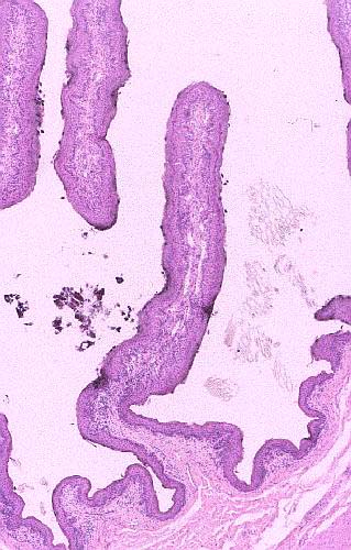
despite the nature of the lining. The image at right is of a single papilla.
It has a core of lamina propria, and no muscularis mucosae. At this magnification
you can see the entire length of this papilla, and the absence of the muscularis
mucosae is more obvious.
Picture Credit: I am indebted to Dr. Mohammed Khalil of
Purdue University's College of Veterinary Medicine for the scanning EM image
here.
 Despite
the presence of keratinized stratified squamous epithelium, a considerable amount
of nutrient absorption occurs in the rumen. Volatile fatty acids produced from
cellulose by the symbiotic bacteria of the tract are absorbed into the blood vessels
in the CT layer.
Despite
the presence of keratinized stratified squamous epithelium, a considerable amount
of nutrient absorption occurs in the rumen. Volatile fatty acids produced from
cellulose by the symbiotic bacteria of the tract are absorbed into the blood vessels
in the CT layer.
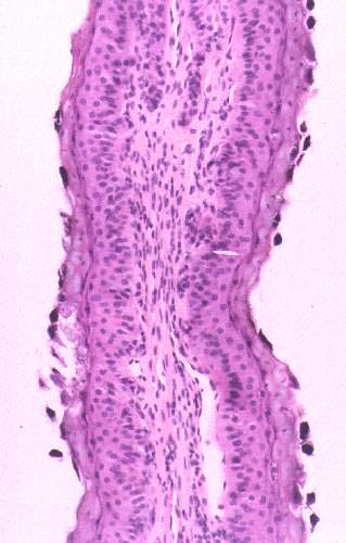
A higher magnification of this area shows more detail, as seen at right: the nature of the epithelium becomes clear. Note that the
keratinization is fairly heavy in this part of the forestomach; it becomes less
so in the deeper parts, but nevertheless keratinization is a feature of the
reticulum and the omasum as well.
This image shows some black flecks close to the epithelium. These
aren't artifact. They are the preserved remains of the ciliated
microorganisms that live in the forestomach and are the actual means
by which the energy contained in cellulose is utilized.
Bovine rumen; H&E stain, paraffin sections, 20x, 40x, and 200x

Close This Window