 The
proventriculus is the glandular portion of the avian compound stomach, and a
rather peculiar organ it is. There's nothing like it in mammals.
  The
proventriculus is the glandular portion of the avian compound stomach, and a
rather peculiar organ it is. There's nothing like it in mammals.
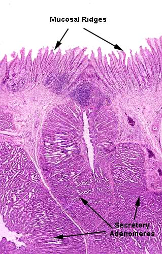
A low power view shows the arrangement of the wall in the proventriculus. The free surface is covered with circular folds of the mucosa, which here appear as finger-like projections. These folds surround a opening into the glandular region below. The surface epithelium on the folds is a simple columnar type, and each fold has a core of lamina propria to support it. These aren't villi, though they might at first glance be mistaken for them. The folds are circular and form a sort of "bull's-eye" arrangement with the central opening into the gland.
The glands proper are in the submucosa; there's a muscularis mucosae between them and the overlying mucosal folds. Each secretory region, or adenomere is a well defined unit. They are separated from each other by a CT investment that outlines and defines the limit of each adenomere. A thin tunica muscularis (not shown) forms the outermost part of the wall in this region of the tract.
This scanning electron micrograph below gives a 3-dimensional view of the adenomere
(SA) and demonstrates how each such secretory region is a separate entity,
clearly demarcated by connective tissue from its neighbors. The tunica muscularis
(TM) of the proventriculus is rather thin, and underlies the adenomeres.
Blood vessels (BV) run through the CT septae. In this striking picture you can see the quasi-globular shape of the secretory adenomere (SAB) and the septations of connective tissue that set it off from adjacent ones. The tunica muscularis (TA) is rather thin, compared to that of the gizzard. Large blood vessels (BV) supply the considerable metabolic needs of this very active organ.
 The
image at right shows the channel that leads from the surface of the proventricular
lumen to the secretory portions of the adenomere. This mid-sagittal section clearly
demonstrates the connection between the source of digestive enzymes and hydrochloric
acid, and the place where they're used.
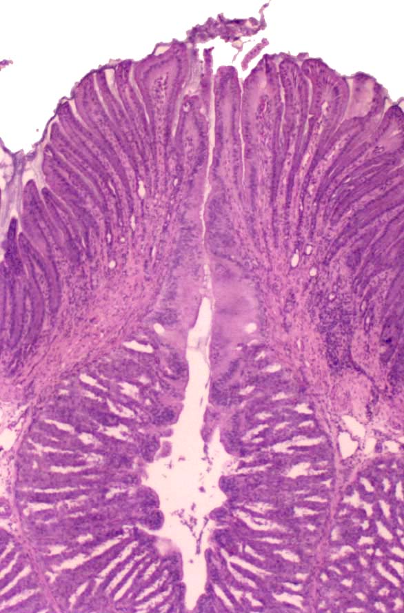
At higher magnification the shape and nature of the mucosal folds or ridges is a little more obvious. These structures are of uniform height, and they have depth; each projects into and out of the plane of the section. Below them can be seen the uppermost part of the underlying adenomere and some of its lumen.
Picture Credit: I am indebted to Dr. Ihab Mahmoud El-Zoghby of the Faculty of Veterinary Medicine, Zagazig University, Egypt, for images #2 and #3. These images are from his doctoral dissertation.
Avian proventriculus; H&E stain, paraffin sections, 20x and 40x; scanning electron microscope preparation, 100x
