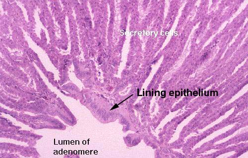
The secretory part of the adenomere consists of radiating arrays of cell plates (a little reminiscent of the physical arrangement of the liver) around a central lumen. The lumen itself is lined with a nonsecretory epithelium of columnar type. Secretions produced in the periphery of the adenomere are discharged into the spaces between the plates, find their way to the center, and out to the surface. Contractions of the muscularis and the stresses of the passage of food hasten the release of the secretions.
The epithelium of the plates is interesting in several ways. It has a peculiar
serrated or "saw toothed" appearance to it. This results from the absence of
junctional complexes at the apices of the cells. Whereas in many epithelial
sheets the cells are held together at the top, these aren't, and in sagging
away from each other the serrated appearance is created. The cells are "simple" in that 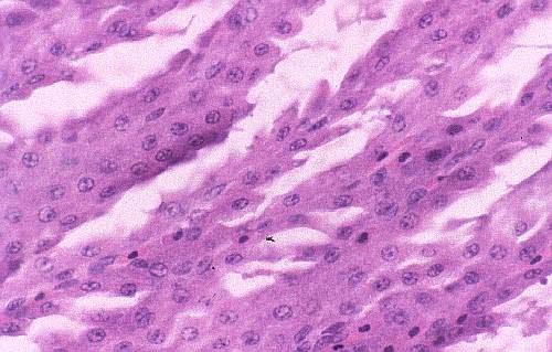 there is only one layer; they sit on a basal lamina, and there's a blood
vessel underneath running through CT. Thus each plate of cells is two cells
thick, with a blood supply in the middle. Notice the erythrocytes: they are
nucleated.
there is only one layer; they sit on a basal lamina, and there's a blood
vessel underneath running through CT. Thus each plate of cells is two cells
thick, with a blood supply in the middle. Notice the erythrocytes: they are
nucleated.
There is only once morphological type of cell in the secretory
areas of the adenomere, but an analysis of the gastric juice from
birds reveals that it has the same basic composition and pH as that
from mammals. Hence it's concluded that the functions of the
mammalian parietal cells and chief cells are carried out by one cell
type in birds.
