VM8054 Veterinary Histology
Ventriculus
Author: Dr. Thomas Caceci
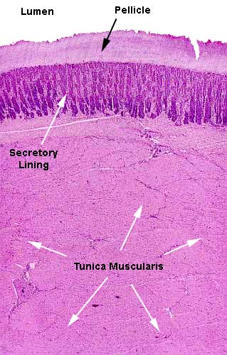
 The enormous thickness of the ventriculus' wall is only suggested
by this low power image. The epithelium and its overlying pellicle
are dwarfed by the immensely thick tunica muscularis; but actually
what you are seeing here is perhaps one third of the real thickness
of the external muscle layer. It was so large it couldn't all have
been put on the microscope slide, let alone on the screen!
The epithelium, as we'll see later, is formed into deep crypt-like
invaginations. The cells produce the pellicle, which is worn away at
the top edge and replaced from below. This image shows only one side
of the ventriculus; a mirror image is present in life.
 The pellicle is nonliving material, of course. It's the secretory
product of the underlying cells, and it's made in a manner similar to
hoof or fingernail material.
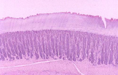
A closer look at the mucosal layer reveals a little more detail. The mucosa is ridged and the ridges and crypts lined with cells. They constitute a simple columnar layer (cuboidal in the deeper parts). This field is coincidentally very similar in layout to the histology of the equine hoof. That's because the job to be done is the same in both cases: a rapidly worn surface composed of cellular product has to be replaced as fast as it's removed.
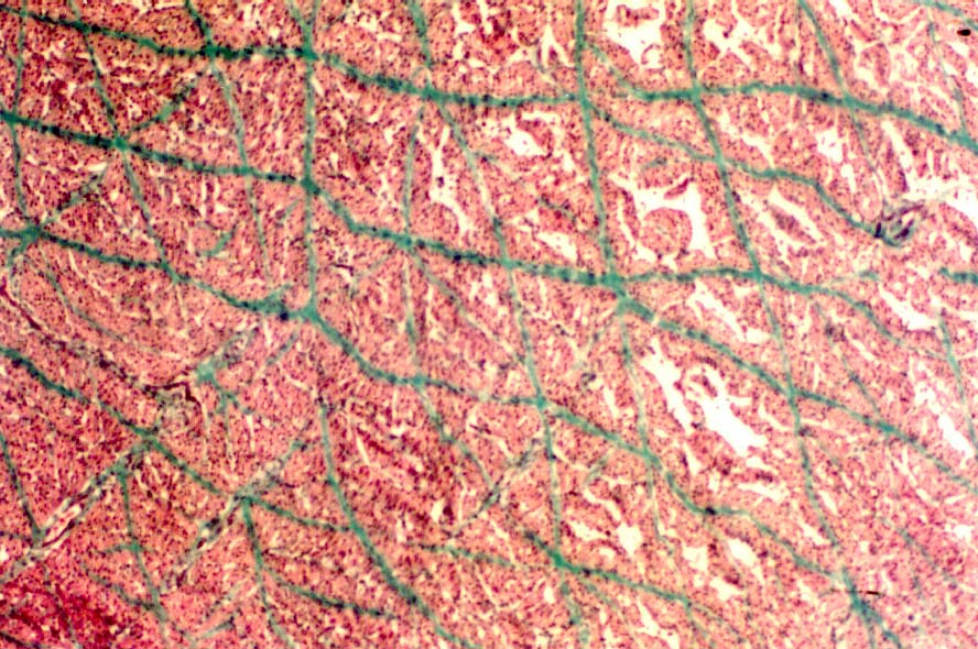
 The
muscular wall of the gizzard is reinforced by collagenous fibers in a regular
array. The hexagonal "meshwork" pattern seen in the image to the left
is this reinforcement. The gizzard grinds constantly; it works against itself
to pulp the food and reduce it to mush. The intercellular collagen network is
extremely stout, much more developed than that of the heart or skeletal muscle.
Anyone who has ever cooked a gizzard will know that a couple of hours' boiling
is needed to reduce it to the point where it can be chewed; and then it falls
apart into neat bundles, along the planes of the collagen reinforcement.
Picture Credit: I am greatly
indebted to Dr. Ihab Mahmoud El-Zoghby of the Faculty of Veterinary Medicine,
Zagazig University, Egypt, for this image. It is from his doctoral dissertation.
Chicken ventriculus; H&E stain, paraffin sections, 20x and
40x; Turkey ventriculus, paraffin section, Masson's stain, 100x

Close This Window
 The enormous thickness of the ventriculus' wall is only suggested
by this low power image. The epithelium and its overlying pellicle
are dwarfed by the immensely thick tunica muscularis; but actually
what you are seeing here is perhaps one third of the real thickness
of the external muscle layer. It was so large it couldn't all have
been put on the microscope slide, let alone on the screen!
 The pellicle is nonliving material, of course. It's the secretory
product of the underlying cells, and it's made in a manner similar to
hoof or fingernail material.
 The
muscular wall of the gizzard is reinforced by collagenous fibers in a regular
array. The hexagonal "meshwork" pattern seen in the image to the left
is this reinforcement. The gizzard grinds constantly; it works against itself
to pulp the food and reduce it to mush. The intercellular collagen network is
extremely stout, much more developed than that of the heart or skeletal muscle.
Anyone who has ever cooked a gizzard will know that a couple of hours' boiling
is needed to reduce it to the point where it can be chewed; and then it falls
apart into neat bundles, along the planes of the collagen reinforcement.