VM8054 Veterinary Histology
Mesobronchus & Air Passages
Author: Dr. Thomas Caceci
 The
mesobronchus is structurally very similar to the mammalian bronchus,
as the name implies. It's lined with TRE and has cartilage and smooth muscle
in its walls. It has no direct function in gas exchange; as with the mammalian
bronchus it's an airway and not a respiratory surface. This image (and the one
below) show a mesobronchus cut in cross section. Notice the presence of hyaline
cartilage plates in the wall.
The
mesobronchus conducts air through the middle of the lung and also to 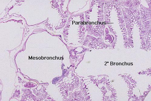 smaller bronchi.
It gives rise to recurrent secondary bronchi, which in their turn give
rise to tertiary bronchi (also called parabronchi). (The secondary
bronchi, as you can see here, have walls made primarily of smooth muscle, and
in a mammal might more properly be termed "bronchioles.")
smaller bronchi.
It gives rise to recurrent secondary bronchi, which in their turn give
rise to tertiary bronchi (also called parabronchi). (The secondary
bronchi, as you can see here, have walls made primarily of smooth muscle, and
in a mammal might more properly be termed "bronchioles.")
The parabronchi coming off the secondaries have a peculiar structure that's difficult to appreciate
in two dimensional sections; the walls of these airways are "scalloped" by the bay-like air vesicles, the place where actual gas exchange takes
place.
Avian lung; H&E stain, paraffin sections, 20x and 20x

Close This Window