Mechanics of Respiration in Birds
Birds ventilate their lungs not by expanding the lungs themselves,
but by expanding the air sacs, a uniquely avian anatomical
feature. The air sacs are balloon-like structures at the "ends" of
the airway system, each with a wall of squamous epithelium. In the
chicken, the "model" species, there are eight such sacs: an unpaired
one in the cervical region; another unpaired one in the clavicular
area; two paired sets (cranial and caudal) in the thorax; and a
paired set in the caudal abdomen.
The key to the system is that distention and compression of the
air sacs, not the lungs, moves air in and out and that at any given
moment air may be moving into and out of the lung and being "parked"
in the air sacs. The lungs are stiff and noncompliant, not at all
like the distensible lungs of mammals. The air sacs act as "bellows" to suck air in and blow it out, and also to hold part of the total
volume.
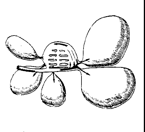
 Here's
the condition at the beginning of each inspiration of the respiratory cycle
(only one side is shown). The cranial end of the system is shown to the left,
and the trachea is shown. Air will enter and leave via the trachea as it does
in mammals. All the air sacs are deflated at this stage. There is "resting" air in the lungs. Each one of a bird's paired lungs has a single large mesobronchus,
essentially the intrapulmonary continuation of the trachea, which runs straight
through it. This is a long straight airway, much like a mammalian bronchus in
construction, which connects the trachea to the caudal air sacs. It also serves
to ventilate the lung directly with part of each incoming breath.
Here's
the condition at the beginning of each inspiration of the respiratory cycle
(only one side is shown). The cranial end of the system is shown to the left,
and the trachea is shown. Air will enter and leave via the trachea as it does
in mammals. All the air sacs are deflated at this stage. There is "resting" air in the lungs. Each one of a bird's paired lungs has a single large mesobronchus,
essentially the intrapulmonary continuation of the trachea, which runs straight
through it. This is a long straight airway, much like a mammalian bronchus in
construction, which connects the trachea to the caudal air sacs. It also serves
to ventilate the lung directly with part of each incoming breath.
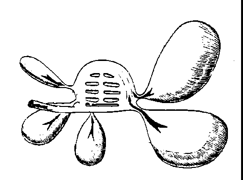
 At
the moment of inspiration, air enters via the trachea and is routed to the mesobronchus.
Half of the incoming air goes directly into the lung, and the other half goes
directly (via the mesobronchus) into the caudal air sacs. The "resting" air that was in the lungs now moves out of them and into the cranial
air sacs. Note the direction of flow indicated by the arrows, and the inflated
state of the air sacs. They're inflated by muscular movements, as the chest
expands and the ribs move ventrally and cranially. This distention pulls air
into the caudal sacs via the inspiratory route, and into the cranial sacs from
the lungs. Gas exchange occurs during this phase of the cycle, as half of the
inspired air moves into the lung directly.
At
the moment of inspiration, air enters via the trachea and is routed to the mesobronchus.
Half of the incoming air goes directly into the lung, and the other half goes
directly (via the mesobronchus) into the caudal air sacs. The "resting" air that was in the lungs now moves out of them and into the cranial
air sacs. Note the direction of flow indicated by the arrows, and the inflated
state of the air sacs. They're inflated by muscular movements, as the chest
expands and the ribs move ventrally and cranially. This distention pulls air
into the caudal sacs via the inspiratory route, and into the cranial sacs from
the lungs. Gas exchange occurs during this phase of the cycle, as half of the
inspired air moves into the lung directly.
 At
the beginning of the exhalation, the air in the caudal air sacs (which are beginning
to deflate) moves into the lungs (this was the "second half" of the
air inspired before, remember). The air that was in the lungs and the cranial
air sacs now moves out via the trachea. This movement results from compression
of the chest and abdomen by muscle movement, and it's the reverse of the previous
step.
At
the beginning of the next inspiration, the situation is as it was in the first
diagram, except that the air which was in the lungs at the beginning has been
replaced by air from the caudal sacs; the cranial sacs are empty, and half of
the air from the first breath is now in the caudal sacs. With
the end of second inhalation, half of the volume of the first breath moves into
the cranial air sacs; it will be expelled via the trachea in the 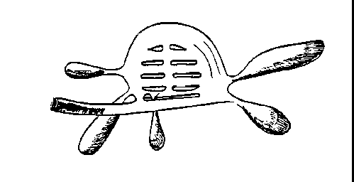 next exhalation.
As before, air moves from the caudal sacs into the lung. The cycle repeats in
this way, with about one third the total volume of the system being moved from
one place to the next through each inspiratory and expiratory phase.
next exhalation.
As before, air moves from the caudal sacs into the lung. The cycle repeats in
this way, with about one third the total volume of the system being moved from
one place to the next through each inspiratory and expiratory phase.
Gas exchange in the lung is continuous throughout the process,
with one half the inspired volume of any given breath being "processed" at any given moment to extract oxygen. This setup
amounts to a "flow through" system, and rather than the in and out
breathing cycle we have, birds respire more or less continuously.
This has great importance because flight demands an enormous amount
of oxygen exchange, far more than is needed for terrestrial
locomotion, and the respiratory system of birds is very efficient at
providing it. 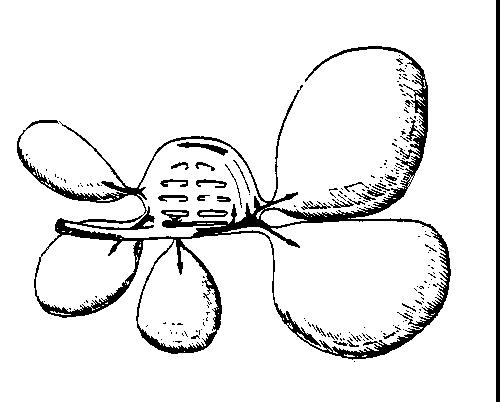 This is due only in part to the "bellows" action of
the air sacs; it depends in great measure on the presence of a
"countercurrent flow" mechanism for gas exchange in the lung proper,
which we'll look at next.
This is due only in part to the "bellows" action of
the air sacs; it depends in great measure on the presence of a
"countercurrent flow" mechanism for gas exchange in the lung proper,
which we'll look at next.
I am greatly indebted to Dr. Charles J. McGrath of
the VMRCVM's Large Animal Clinical Sciences Department for the
illustrations used above.

Close This Window