VM8054 Veterinary Histology
Fimbriae
Author: Dr. Thomas Caceci
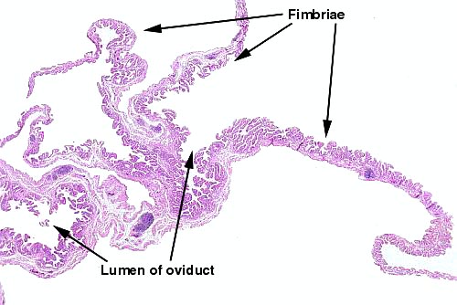
 The oviduct of birds, like that of mammals, has an expanded upper end, to catch the released egg. The size of the infundibulum in birds is proportional to the size of the egg. In this image, the upper end of the tract is at the right side of the screen; and the opening is expanded to include long fingerlike fimbriae.
This is a section through the infundibular part of the oviduct;
the fimbriae are the long, slender, finger-like projections to
the upper right. Each is covered with a ciliated simple columnar
epithelium. The beating of the cilia is important in moving the egg
into the funnel shaped upper end of the tube.
The infundibulum also has a secretory function. It produces the first of the egg coats, the chalazae. These are the whitish string-like structures on either side of the yolk, that keep the embryo in proper position during development.
 The ciliation on the mucosal lining of the fimbriae is very heavy,
as you can see here.
The gross arrangement of the fimbriae and the
beating of the ciliated epithelium creates a vortex to pull in the
egg. The ciliation here is so extensive that it was from this organ
that these organelles were first reported, in the early 19th Century.
Note the simple nature of the epithelium and the well vascularized
CT of the underlying lamina propria.
Oviduct, chicken; H&E stain, paraffin sections, 20x and
200x

Close This Window
 The oviduct of birds, like that of mammals, has an expanded upper end, to catch the released egg. The size of the infundibulum in birds is proportional to the size of the egg. In this image, the upper end of the tract is at the right side of the screen; and the opening is expanded to include long fingerlike fimbriae.
