Structural Classification of Exocrine Organs
"Epithelial Organs"
Most glands (and some other organs) are spoken of as being "epithelial organs." While it's conventional to
this
nomenclature, technically it's incorrect, since it implies that the organ contains only
epithelium, and no other tissue type. Obviously this can't be the case—an
organ is, by definition, "a collection of tissues that are integrated to
carry out certain functions"—so what is really meant is that they
are composed principally of epithelial cells.
Glands
Exocrine glands are those with ducts to the outside world,
and whose secretions are carried through those ducts. While the bulk of the gland is made of secretory
or glandular epithelium, there will always be a fair amount of "covering
and lining" type as part of the organ. The neat and tidy "covering and lining" classification is inapplicable to the actual secretory areas, but it applies very well to the ducts that carry the gland's secretions. .
Exocrine organs as a whole can be classified structurally, however,
based on the nature of the ductwork. The two broadest categories are simple glands and compound glands.
Simple glands have undivided
ducts; compound glands are those whose ducts divide at least once. It's important to remember that the term "compound" applies only to the ductwork, not to the secretory regions.
The second criterion used in structural classification of exocrine glands is the shape of their secretory
regions, of which more will be said presently.
Simple Exocrine Glands
A simple exocrine gland is one which has an unbranched duct. i.e.,
a straight tube. Let's look at some examples.
Simple Tubular and Simple Coiled Tubular Glands
 The least complicated form of simple gland is one in which the secretory region, as well as the duct, is straight;
such a gland
would be classified structurally as "simple tubular." A variant of
this form is one in which the secretory region is wound into a coil; it's
still a simple tube, without branching. It's classified as "simple coiled
tubular." The difference is that between a garden hose laid out straight
and one wound up on a hose reel.
The least complicated form of simple gland is one in which the secretory region, as well as the duct, is straight;
such a gland
would be classified structurally as "simple tubular." A variant of
this form is one in which the secretory region is wound into a coil; it's
still a simple tube, without branching. It's classified as "simple coiled
tubular." The difference is that between a garden hose laid out straight
and one wound up on a hose reel.
Simple tubular glands aren't all that common in mammals: about the only easily found example is
the large intestine. Examine slide 126. The luminal surface of the large intestine
has openings into deep "intestinal glands" (more properly called "intestinal crypts"). These deep, straight invaginations, much like what would result if
you were to push your finger down into a block of modeling clay, are seen in this image. They're
lined with epithelium, and they do in fact have some secretory cells (goblet cells) in them. The secretions are discharged via an opening to the
surface. This slide is stained with Alcian Blue, which highlights the carbohydrate content of the goblet cells, making them appear as blue dots against the H&E background stain.
I have to be honest and say that calling these "glands" has always bothered me. Yes, they're secretory, but the fact is that they don't quite fit the concept of a solid mass of cells that have a secretory function. Nor is it consistent with the concept of the goblet cell as a "unicellular gland," as it's often called.
 The next most complicated form of simple gland is the "simple coiled
tubular type." The sweat glands of the skin are this type, and can be
seen in slides 11, 12, and 35. You will see sweat glands near the bases
of the hairs on these slides.
The next most complicated form of simple gland is the "simple coiled
tubular type." The sweat glands of the skin are this type, and can be
seen in slides 11, 12, and 35. You will see sweat glands near the bases
of the hairs on these slides.
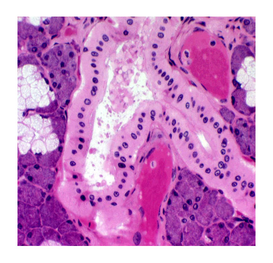
This is a tough thing to visualize in 3-D from a section, but imagine a garden hose, lying in a tangle on the ground. Now pull one end of the hose up: that's the duct. The still-tangled part is the secretory region. Now, if you visualize passing a plane through that tangle, you'd be looking into the cut sections, which will appear as small round
to oval profiles. If the plane of the section passes through the center of the duct (and if the duct is straight, which it usually isn't) you'd see that connected to them. In the image at right (from slide 35) you see the "cut ends" of the secretory areas and the beginning of the duct system. If you were to inject a dye into any portion of this gland it would reach all the other parts of it, because it's one continuous system.
Simple Branched Tubular Glands
 A gland can have a simple duct and a subdivided (or "branched") secretory region.
Since the duct doesn't divide, such an arrangement is still classified as "simple." The designation for a gland like this—undivided duct, divided secretory region—is "simple branched tubular." An example of this is found in
the of glands in the lumen of the stomach. You will see this type of gland on slide 131. The lining of the stomach
(like that of the large intestine) has glandular properties. In this case,
the secretory portions of the glands (in the deep regions) are branched,
and two or more such secretory areas open into a single common duct, which
leads the secretions to the surface. The gland as a whole is shaped like an inverted letter Y with the stem as the neck. Note the nomenclature carefully. This is a simple gland. Don't make the mistake of confusing "compound" with "branched." The former term refers to ducts, the latter
to secretory regions.
A gland can have a simple duct and a subdivided (or "branched") secretory region.
Since the duct doesn't divide, such an arrangement is still classified as "simple." The designation for a gland like this—undivided duct, divided secretory region—is "simple branched tubular." An example of this is found in
the of glands in the lumen of the stomach. You will see this type of gland on slide 131. The lining of the stomach
(like that of the large intestine) has glandular properties. In this case,
the secretory portions of the glands (in the deep regions) are branched,
and two or more such secretory areas open into a single common duct, which
leads the secretions to the surface. The gland as a whole is shaped like an inverted letter Y with the stem as the neck. Note the nomenclature carefully. This is a simple gland. Don't make the mistake of confusing "compound" with "branched." The former term refers to ducts, the latter
to secretory regions.
Compound Glands
These are exocrine glands in which the duct system is divided, although
the secretory portion may or may not be. The secretory portions may take
different shapes, and this is part of the classification system, too. Any gland whose DUCT is divided is "compound."
Compound Tubular Glands
Such a gland has a divided duct and a secretory region that's tubular in shape.
One example is the large submucosal gland of the duodenum. These glands, which you'll see on slide 40,
lie in the region between the epithelial lining and the muscular outer wall
of the first portion of the intestines.
Other Secretory Region Shapes
 The exocrine organs considered thus far have all been classified partly
on the shape of the secretory region, as well as the duct; that's why they
are all "something-or-other-TUBULAR." There are (of course!) other shapes
for secretory regions.
The exocrine organs considered thus far have all been classified partly
on the shape of the secretory region, as well as the duct; that's why they
are all "something-or-other-TUBULAR." There are (of course!) other shapes
for secretory regions.
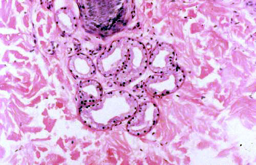
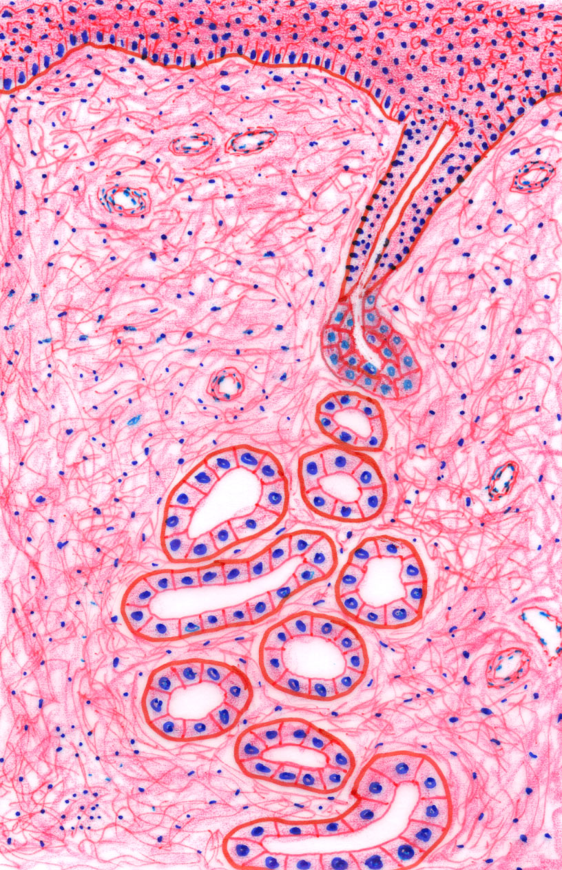
 An acinar or alveolar secretory region
is one whose three-dimensional shape is something like a grape or an olive. The duct may be
simple, in which case you would classify it as a "simple branched acinar
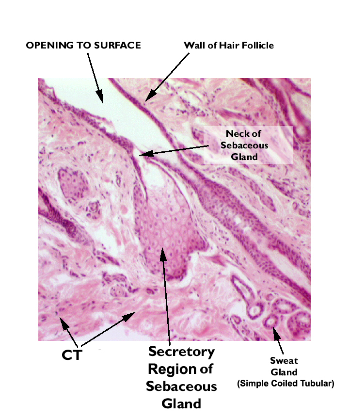
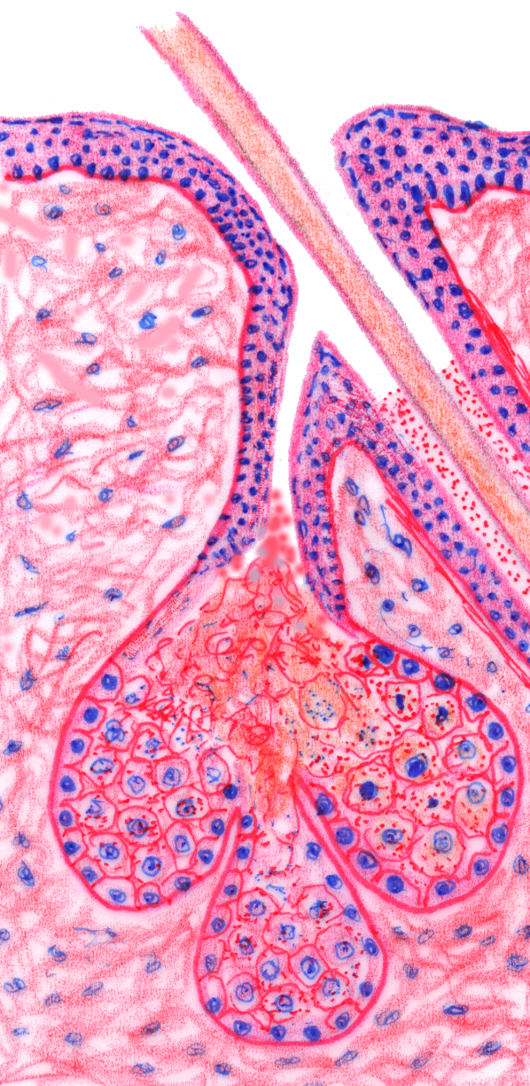
(or alveolar) gland." The only example of such a gland is the sebaceous
gland. You will find some on slides 11 and 12, and they should look like the one on the left, which is from slide 11. They're recognizable by their bag-like secretory
areas, all of them emptying into a common short duct, which
in turn leads into the space through which the hair runs. The sketch to the right, which may make the layout of this type of gland a little more comprehensible, shows at least three of these bag-like secretory regions, all of them opening into the same duct, which is very short and discharges the combined secretions into the hair shaft tunnel. Hence the "branched" and "simple" designation. We'll discuss sebaceous glands in more detail in the laboratory exercise on the integumentary system.
An acinar or alveolar secretory region
is one whose three-dimensional shape is something like a grape or an olive. The duct may be
simple, in which case you would classify it as a "simple branched acinar
(or alveolar) gland." The only example of such a gland is the sebaceous
gland. You will find some on slides 11 and 12, and they should look like the one on the left, which is from slide 11. They're recognizable by their bag-like secretory
areas, all of them emptying into a common short duct, which
in turn leads into the space through which the hair runs. The sketch to the right, which may make the layout of this type of gland a little more comprehensible, shows at least three of these bag-like secretory regions, all of them opening into the same duct, which is very short and discharges the combined secretions into the hair shaft tunnel. Hence the "branched" and "simple" designation. We'll discuss sebaceous glands in more detail in the laboratory exercise on the integumentary system.
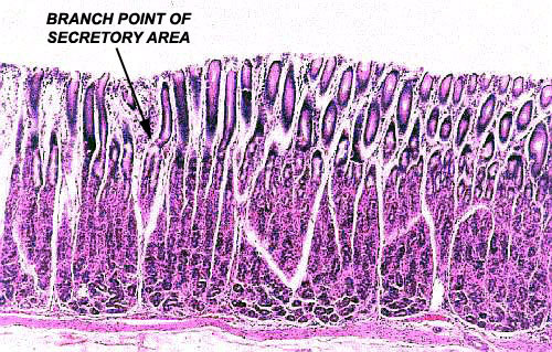
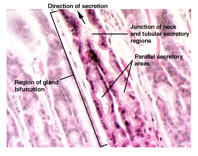
 Compound acinar (or alveolar) glands are common. Most exocrine glands have this structure. The best
examples are salivary glands, and slide 28 bears a good example
of the "compound acinar" gland structure. The secretory regions, which are quite distinct
from the ducts, are of the acinar shape, and when cut in cross section,
show a round profile. Notice that the ducts, which are "covering and lining" epithelium, are usually composed of simple cuboidal; in some places, where
the ducts are very large, it may become stratified cuboidal.
Compound acinar (or alveolar) glands are common. Most exocrine glands have this structure. The best
examples are salivary glands, and slide 28 bears a good example
of the "compound acinar" gland structure. The secretory regions, which are quite distinct
from the ducts, are of the acinar shape, and when cut in cross section,
show a round profile. Notice that the ducts, which are "covering and lining" epithelium, are usually composed of simple cuboidal; in some places, where
the ducts are very large, it may become stratified cuboidal.
Notice that in the image shown here from slide 28, there is more than one duct profile visible in the same plane of the section. That's significant. Remember: a gland (and of course its duct system) has three dimensionality, even though you're seeing it in two dimensions. Moreover, the duct system is arranged like a tree trunk and branches. No matter how small a twig may be, ultimately it's connected to the trunk; so it is with ducts: small ones drain into large ones, and those into still larger ones, and eventually all the secretions are collected into one (or two) main outflow ducts, the "trunk" of the "tree." THEREFORE, if you can actually see a branch point—for that matter, if you can see more than one duct profile at all—in the same section plane, the gland MUST be "compound," because all the ducts are connected. If you passed a plane through the branches of a tree, you'd see "limb profiles" of all sizes, but you know based on the gross anatomy of trees that they're all part of one system, right?
The final
classification is also seen on slide 28. This is the "compound tubuloacinar" (or tubuloalveolar)
shape. In this sort of gland, the secretory regions often contain straight (i.e. tubular) portions which
are connected to round or oval (i.e. acinar or alveolar) areas.
As with any other exocrine glands, the ductwork proper can also be structurally classified using the
"covering and lining" criteria, independent of the secretory regions, which (by definition) aren't "C&L" epithelium.
Endocrine Glands
Endocrine glands are also epithelial organs. They are distinguished
by being "ductless" and therefore are not structurally categorized, as
are exocrine glands. We'll deal with these in much more detail in another exercise. Endocrine glands are predominantly cellular aggregations, with relatively
little connective tissue, though of course there is always some. Blood vessels are present, typically running through thin
regions of CT. Since endocrine glands release their products into the blood, a good deal of vascularization is expected.
Close This Window and Return to Exercise