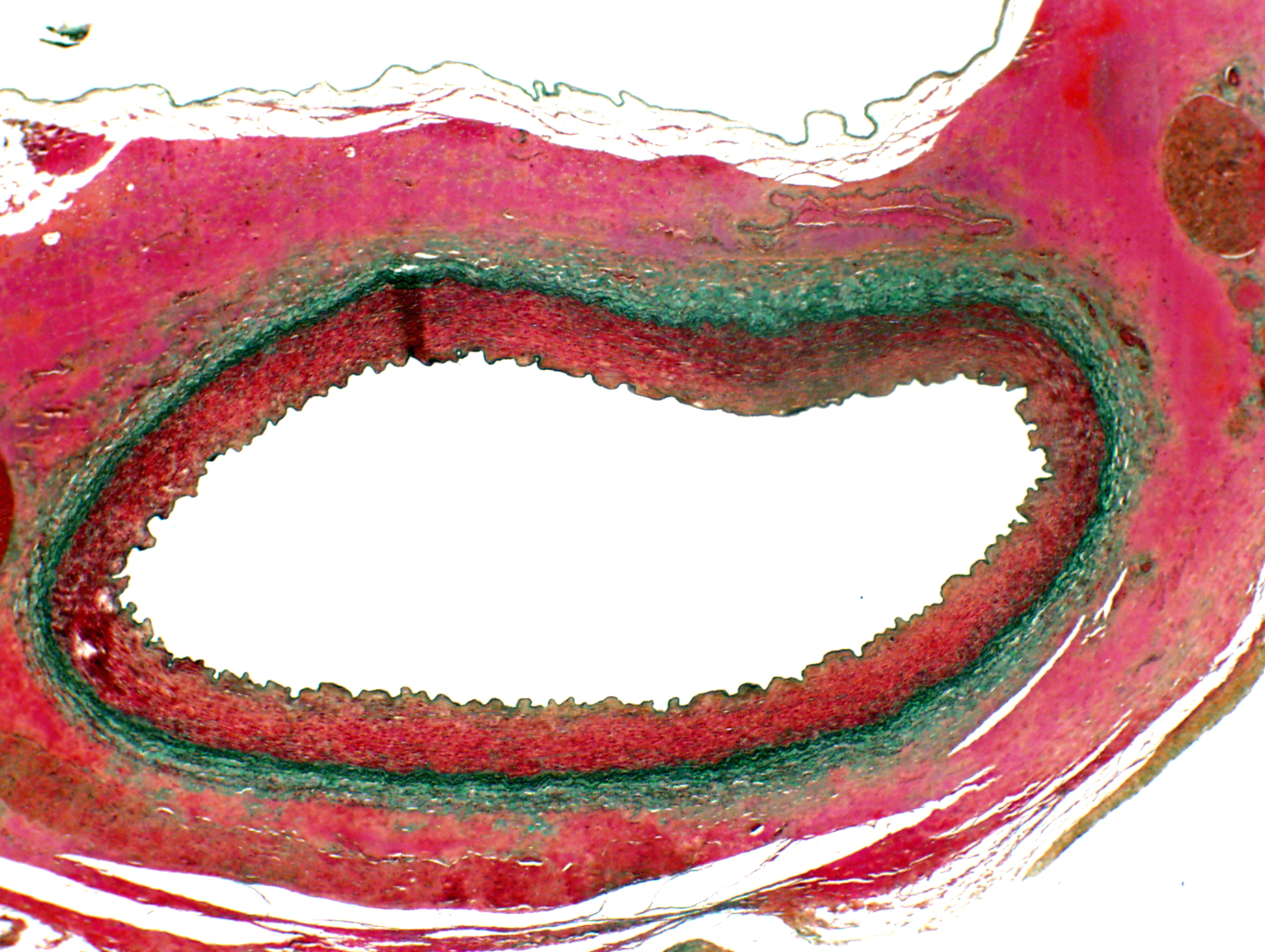
This stain combination is named for Frederick Verhoeff (1874-1968), an American ophthalmologist, and C.L. Pierre Masson (1880-1959), a Canadian pathologist. In it the Masson's Trichrome stain, and the Verhoeff stain, both routines for connective tissue, have been combined. The Masson stain colors collagen green and muscle red; the Verhoeff stain colors elastic fibers black. Using a combination method like this one makes it possible to provide a lot of detail about how an organ is built. Arteries are supposed to resist bursting under pressure. Black elastic fibers are interspersed throughout the muscle to provide springiness and resilience; the green collagen fibers confer resistance to overstretch.
Component-specific staining routines like this one are a very valuable tool in determining the source of pathologies. The image here is from a normal artery; one from an animal with a chronic condition interfering with connective tissue synthesis would look very different with the combined stain, but probably not with a general stain like H&E. Either routine could provide important information, but employing both would permit a much more specific diagnosis to be made.
Ringed seal (Phoca hispida) renal artery: Verhoeff/Masson Stain, 200x and 400x
| H&E | PAS | Masson's CT Stain | Verhoeff-van Gieson | Verhoeff-Masson | Mallory's CT Stain | Golgi Stain|
| Cresyl Violet | Cresyl Violet-Luxol Fast Blue | Kluver-Barrera | Fontana-Masson | Prussian Blue | Toluidine Blue|
|Osmium Tetroxide | Oil Red O | Sudan Black | Fluorescent & Enzymatic Tagging |