The Three Tunics of the Eye
The wall of the eyeball is made up of three coats, or tunics,
one inside the other. From outermost to innermost, these are the corneoscleral
tunic, the uveal tunic and the retinal
tunic.
The corneoscleral tunic's functions are chiefly mechanical
and optical. The tough, opaque, collagenous portion, the sclera, is the
so-called "white of the eyes"; this is the protective outermost boundary
that encloses all the others. It is the site of insertion of the muscles that
rotate the eye, as well as the route for entry and exit of blood vessels; and
it merges with the cornea at its most cranial portion. The cornea is
the chief light-refracting structure, the first of the
two lenses in the optical system.
 In
the image at left, the three tunics are peeled away to show their relationship
to each other. Note also that there are two blood supplies: one for the
extensive plexus of the choroid portion of the uveal tunic,
and an independent supply to the retina. The embryonic
hyaloid artery and vein are the source of the retina's vasculature. The distal
ends of these vessels degenerate as the lens and vitreous body mature; and the
remaining proximal portions are enclosed in the forming optic nerve.
In
the image at left, the three tunics are peeled away to show their relationship
to each other. Note also that there are two blood supplies: one for the
extensive plexus of the choroid portion of the uveal tunic,
and an independent supply to the retina. The embryonic
hyaloid artery and vein are the source of the retina's vasculature. The distal
ends of these vessels degenerate as the lens and vitreous body mature; and the
remaining proximal portions are enclosed in the forming optic nerve.
The uveal tunic has supportive and nutritive functions for
the globe; it also produces pigments to minimize internal reflections and some
of its cells are of vital importance in the maintenance of the light-sensitive
parts of the retina. The uveal tunic also gives rise to the muscles which control
focusing, and to the iris, the muscular sphincter whose variable diameter
controls the amount of light admitted to the inner portion of the eye.
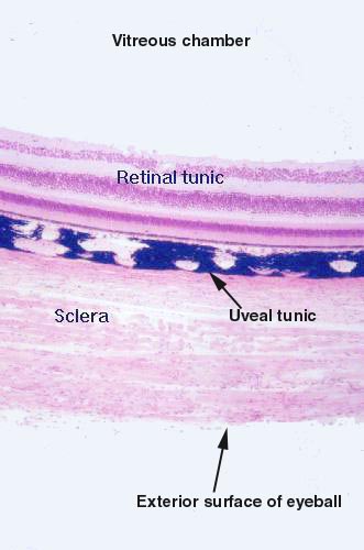 This
color image from an actual specimen shows the relationships of the three tunics
to one another quite well. The uveal tunic is heavily pigmented with melanin,
produced locally to dampen internal reflections and increase acuity of vision.
There are numerous blood channels and lymphatic vessel in it, visible here as
breaks in the melanin.
This
color image from an actual specimen shows the relationships of the three tunics
to one another quite well. The uveal tunic is heavily pigmented with melanin,
produced locally to dampen internal reflections and increase acuity of vision.
There are numerous blood channels and lymphatic vessel in it, visible here as
breaks in the melanin.
Click Back to return
Or Go To:
Main Page | Corneoscleral Tunic
| Uveal Tunic | Retinal Tunic
| Physiology of Vision | CNS Processing
of Visual Signals