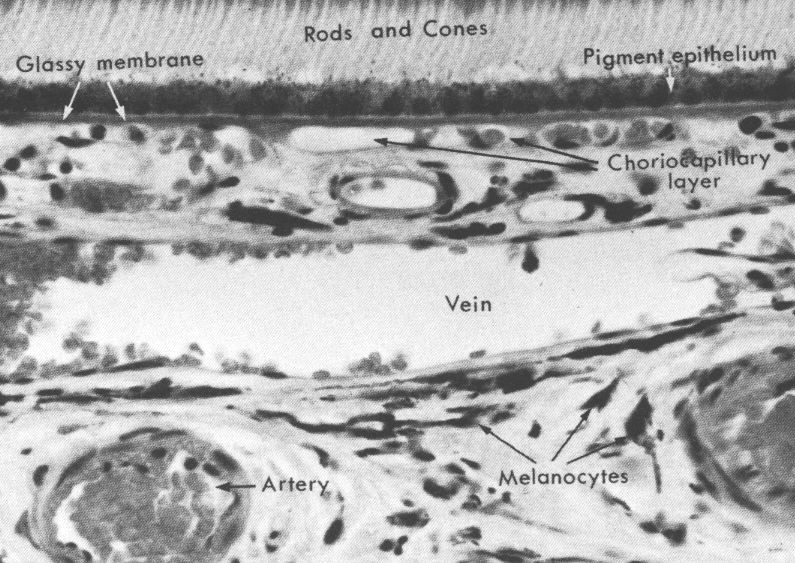
 The choroid is the highly pigmented and vascular layer of the
uveal tunic that underlies the eyeball's surface. It provides nutritive
support, and contains numerous blood vessels and large lymphatic channels.
The choroid is the highly pigmented and vascular layer of the
uveal tunic that underlies the eyeball's surface. It provides nutritive
support, and contains numerous blood vessels and large lymphatic channels.
The uvea is the middle tunic, located between the innermost retina and the outermost corneoscleral tunics.
The uvea has many functions, and in some respects it is the most complex of the eye's tunics. One major function is to provide for most of the eyeball's blood supply and lymphatic drainage (its embryonic origins from mesoderm insure that it forms blood vessels in development). But the uveal tunic is also the location of the mechanism for accommodation, and of control of light influx.
Structurally the uveal tunic has three subdivisions. These are the choroid, the heavily vascular layer which comprises most of it; the iris, the variable-diameter sphincter which controls the amount of light entering the eye; and the ciliary body, a mass of smooth muscle which controls the accomodation mechanism and from which the lens is suspended. The ciliary body also has secretory function; its ciliary processes are the site of production of aqueous humor.
The choroid is the highly pigmented and vascular layer of the
uveal tunic that underlies the eyeball's surface. It provides nutritive
support, and contains numerous blood vessels and large lymphatic channels.
The choroid is also very heavily pigmented in most animals. The pigment is melanin, and melanocytes are found in it. Together with the lipofuscin and melanin in the pigment cell layer of the retina, it acts as a "light trap" in the same way the black matte finish of the inside of a camera does. Absorbing stray light minimizes internal reflections and increases contrast, thus increasing the acuity of vision. If this layer lacks pigmentation, light passing through the retina can be reflected from the inner surface of the sclera, into the light-sensitive rods and cones. This seriously impairs the precision with which the image can be formed.
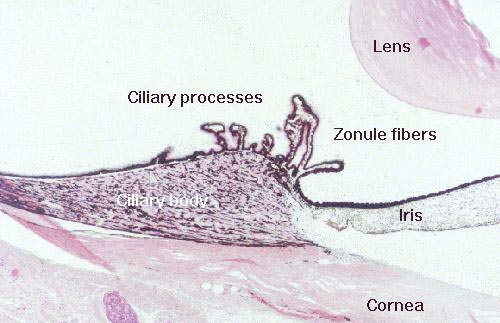 The
ciliary body is an anterior extension and expansion of the uveal tunic, located
in the angle between the end of the cornea and the lens. Most of the ciliary
body is composed of smooth muscle whose contractions work
the lens accommodation mechanism via the tension imposed on the zonule fibers.
Like other structures in this important region (the limbus or corneo-iridial
angle), which is shown in the image to the right, the ciliary body runs radially
around the anterior part of the eye.
The
ciliary body is an anterior extension and expansion of the uveal tunic, located
in the angle between the end of the cornea and the lens. Most of the ciliary
body is composed of smooth muscle whose contractions work
the lens accommodation mechanism via the tension imposed on the zonule fibers.
Like other structures in this important region (the limbus or corneo-iridial
angle), which is shown in the image to the right, the ciliary body runs radially
around the anterior part of the eye.
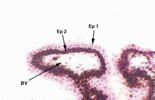 In
addition to its role in the mechanism of accommodation,
the ciliary body is a secretory structure. The inner surface of the ciliary
body is lined on its inner surface with a double layer of cells, the ciliary
epithelium. The outer of these two layers is heavily pigmented, the inner
one is not.
In
addition to its role in the mechanism of accommodation,
the ciliary body is a secretory structure. The inner surface of the ciliary
body is lined on its inner surface with a double layer of cells, the ciliary
epithelium. The outer of these two layers is heavily pigmented, the inner
one is not.
This epithelium is the site of production of the fluid which fills the anterior and posterior chambers of the eye, the aqueous humor. The balance between production and drainage of aqueous humor is the means by which the normal intraocular tension is maintained, and is essential in maintaining the eyeball's normal size and shape.
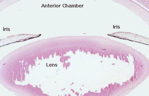
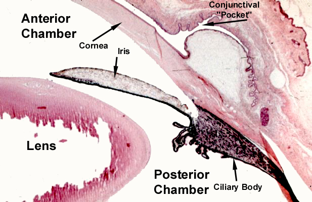 The
iris is the point at which light rays passing through the cornea enter the eye.
The function of the iris is exactly the same as that of the "iris diaphragm"
in the lens of a camera. It contains radially disposed smooth muscle fibers
and a circumferential band of smooth muscle fibers as well. Stimulation of these
opposing muscles controls the amount of light admitted. Contraction of the radial-oriented
fibers opens the pupil, admitting more light; contraction of the circumferential
sphincter muscles reduces the pupillary diameter. These adjustments are made
The
iris is the point at which light rays passing through the cornea enter the eye.
The function of the iris is exactly the same as that of the "iris diaphragm"
in the lens of a camera. It contains radially disposed smooth muscle fibers
and a circumferential band of smooth muscle fibers as well. Stimulation of these
opposing muscles controls the amount of light admitted. Contraction of the radial-oriented
fibers opens the pupil, admitting more light; contraction of the circumferential
sphincter muscles reduces the pupillary diameter. These adjustments are made
 automatically by input from the sympathetic and parasympathetic divisions of
the central nervous system. As is the case with the ciliary body, the iris is
lined on its inner face with an epithelium derived from the non-sensitive portion
of the retina. This is usually pigmented. The bulk of the iris' tissue, from
the uveal tunic, contains some melanocytes and numerous blood vessels. Together
with the pigmentation of the inner layer, the color of the iris as seen from
the external side will vary considerably as a result. The patterns of color
are hereditary in most mammals.
automatically by input from the sympathetic and parasympathetic divisions of
the central nervous system. As is the case with the ciliary body, the iris is
lined on its inner face with an epithelium derived from the non-sensitive portion
of the retina. This is usually pigmented. The bulk of the iris' tissue, from
the uveal tunic, contains some melanocytes and numerous blood vessels. Together
with the pigmentation of the inner layer, the color of the iris as seen from
the external side will vary considerably as a result. The patterns of color
are hereditary in most mammals.
Click Back to return
Or Go To:
Main Page
| Corneoscleral Tunic | Uveal Tunic | Retinal Tunic | Physiology of Vision | CNS Processing of Visual Signals