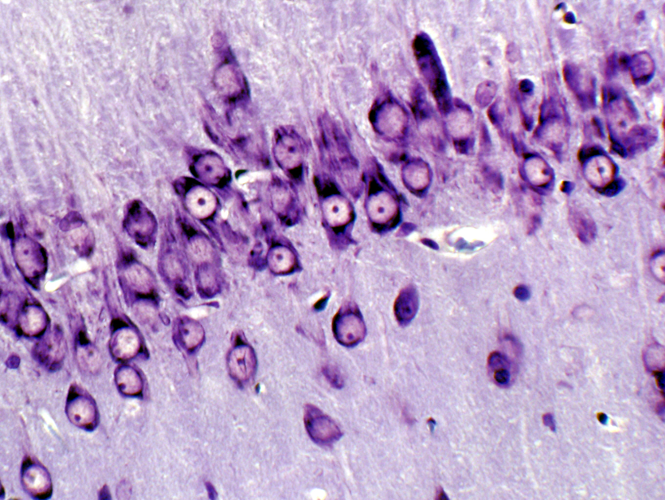
The cresyl violet stain is widely used for nervous tissue. Since it's a basic stain it binds readily to the acidic components of the neuronal cytoplasm, especially to the RNA rich ribosomes, which are present in large numbers in neurons. It binds also to nuclei and nucleoli of the cells in this section. This image is of some large neurons in the cerebellum. If you look carefully at the individual cells you'll notice that some regions within these neurons have taken up the stain even more strongly than others. These areas are the aggregations of rough ER called "Nissl bodies," which will be discussed at greater length in the exercise on organelles. They have so my RNA that they appear almost black. Cresyl violet is probably the third most common stain used in preparing histological sections, and it's often used in conjunction with other stains with an affinity for lipids (such as Luxol Fast Blue).
| H&E | PAS | Masson's CT Stain | Verhoeff-van Gieson | Verhoeff-Masson | Mallory's CT Stain | Golgi Stain|
| Cresyl Violet | Cresyl Violet-Luxol Fast Blue | Kluver-Barrera | Fontana-Masson | Prussian Blue | Toluidine Blue|
|Osmium Tetroxide | Oil Red O | Sudan Black | Fluorescent & Enzymatic Tagging |
