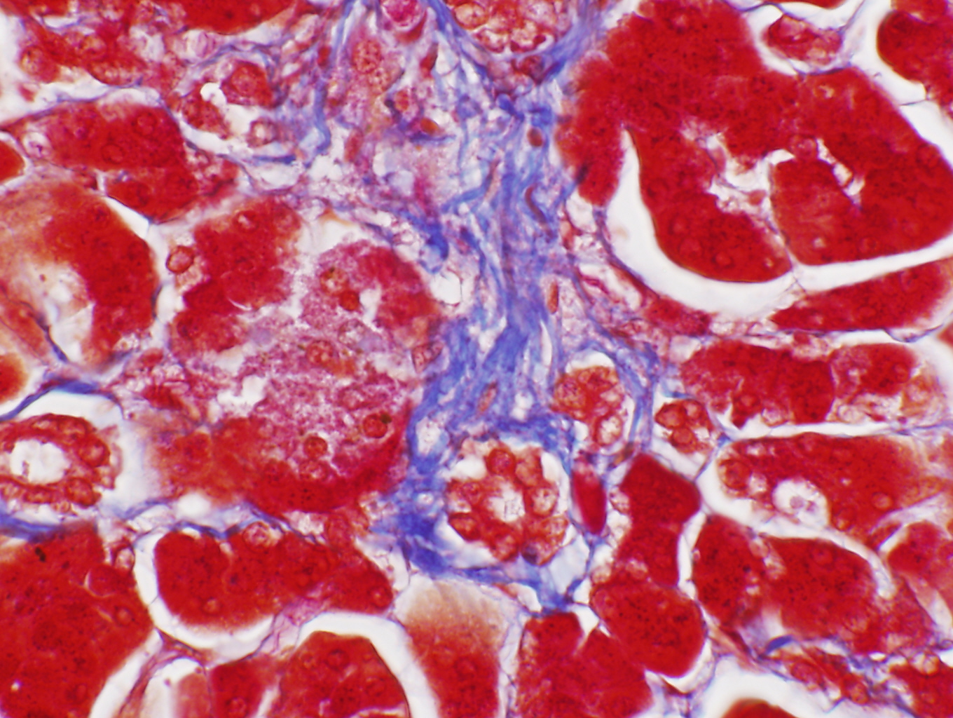
This stain was developed by Dr. Frank Burr Mallory (1862-1941), an American physician and pathologist. It's one of many he invented and it's one of many routines used to identify connective tissue. The example here is from a section of the pancreas. The CT stroma (supporting elements) of this organ are highlighted in blue using this stain, and the parenchymal tissue (i.e., the functional secretory cells) is stained in red. Mallory's CT stain is very widely used and many variations on it exist.
This slide is valuable not only as a demonstration of the method, but as an illustration of just how differently the same organ appears when stained differently. Compare this to the image shown in Exercise 3 as an example of "Basophilia," which is also made from a sample of pancreas. The vast difference in the appearance illustrates a key point: when using stains to highlight chemical components or details of a tissue, you will inevitably trade morphology for specificity. That is, what we think of as the "normal" appearance of a given specimen—it's appearance in an H&E stain—is lost, in exchange for specific information about tissue or organ construction. That's why it's important to use more than one routine to extract the maximum amount of information. One stain is almost never enough to tell you everything you may want to know.
| H&E | PAS | Masson's CT Stain | Verhoeff-van Gieson | Verhoeff-Masson | Mallory's CT Stain | Golgi Stain|
| Cresyl Violet | Cresyl Violet-Luxol Fast Blue | Kluver-Barrera | Fontana-Masson | Prussian Blue | Toluidine Blue|
|Osmium Tetroxide | Oil Red O | Sudan Black | Fluorescent & Enzymatic Tagging |
