Osmium Tetroxide Stain for Lipids
 Osmium tetroxide (OsO4 , structural formula shown at left) is today used more as a fixative for electron microscopy
than as a stain, but it is still one of the best methods for revealing
lipids and lipid-rich structures in the light microscope. The use of OsO4 as a tissue fixative actually goes back to the mid-19th Century. It binds at double bonds of unsaturated lipids and imparts a dense brownish or black color. The chemical reaction that occurs is reduction of the oxide, and deposition of metallic osmium in the tissue.
Osmium tetroxide (OsO4 , structural formula shown at left) is today used more as a fixative for electron microscopy
than as a stain, but it is still one of the best methods for revealing
lipids and lipid-rich structures in the light microscope. The use of OsO4 as a tissue fixative actually goes back to the mid-19th Century. It binds at double bonds of unsaturated lipids and imparts a dense brownish or black color. The chemical reaction that occurs is reduction of the oxide, and deposition of metallic osmium in the tissue.
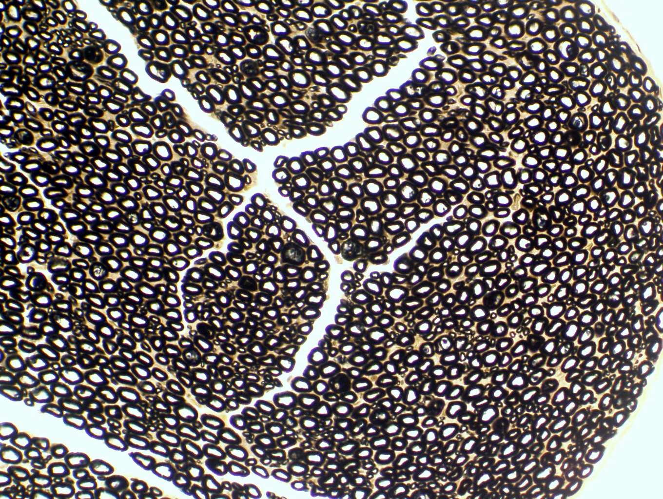
At right is an example: The low magnification image is a cross section of a peripheral nerve. The black circles are individual axons with their coating of lipid-rich myelin. Myelin is a wrapping of extruded plasma membranes of special cells in the peripheral nervous system (see Exercise 9). Because it has such a large concentration of lipid, myelin binds a great deal of the stain. The axon coverings appear blue-black as a result, very easily seen in the high-magnification image provided. Notice also that between the heavily stained axon coverings, there's very little stain reaction: that's because there's connective tissue in these spaces, and CT has virtually no lipid in it. Much less osmium gets deposited in these regions.
In this preparation you see the effects of this
stain: the lipid-rich myelin sheath of nerve fibers is heavily colored.
Peripheral nerve: 100x and 400x
| H&E | PAS | Masson's CT Stain | Verhoeff-van Gieson | Verhoeff-Masson | Mallory's CT Stain | Golgi Stain|
| Cresyl Violet | Cresyl Violet-Luxol Fast Blue | Kluver-Barrera | Fontana-Masson | Prussian Blue | Toluidine Blue|
|Osmium Tetroxide | Oil Red O | Sudan Black | Fluorescent & Enzymatic Tagging |

Close this Window