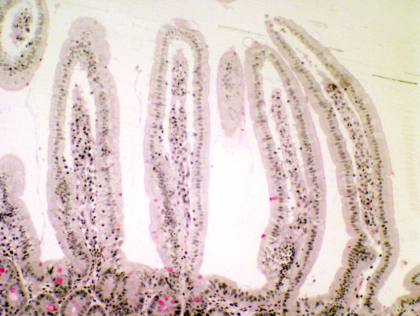
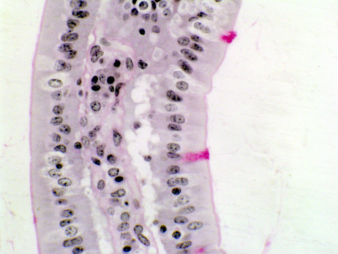
This is an example of the periodic acid-Schiff reaction. These images show villi in the jejunum of a mouse; the right hand rollover image shows the PAS positive cells at higher magnification, and the villi cut longitudinally (as at left) and in cross section as well.
The bright magenta objects in the field are the goblet cells of the epithelial sheet, which respond positively to a PAS stain, which is specific for carbohydrates. In this method, glycol groups are converted to reactive aldehydes by the action of the acid; and the Schiff's Reagent colors them a bright magenta. Polysaccharides are very strongly stained, and since the goblet cells are filled with mucin, they take up the stain preferentially. The rest of the sample has been given a "background" stain of H&E to delineate cell architecture. The PAS reaction is the second most commonly used stain (after H&E) in diagnostic work. The distribution of carbohydrates is usually an important clue to the nature of a tissue and the organs of which it's a part.
Mouse jejunum; PAS stain, 1.5 micron plastic section, 100x and 400x| H&E | PAS | Masson's CT Stain | Verhoeff-van Gieson | Verhoeff-Masson | Mallory's CT Stain | Golgi Stain|
| Cresyl Violet | Cresyl Violet-Luxol Fast Blue | Kluver-Barrera | Fontana-Masson | Prussian Blue | Toluidine Blue|
|Osmium Tetroxide | Oil Red O | Sudan Black | Fluorescent & Enzymatic Tagging |
