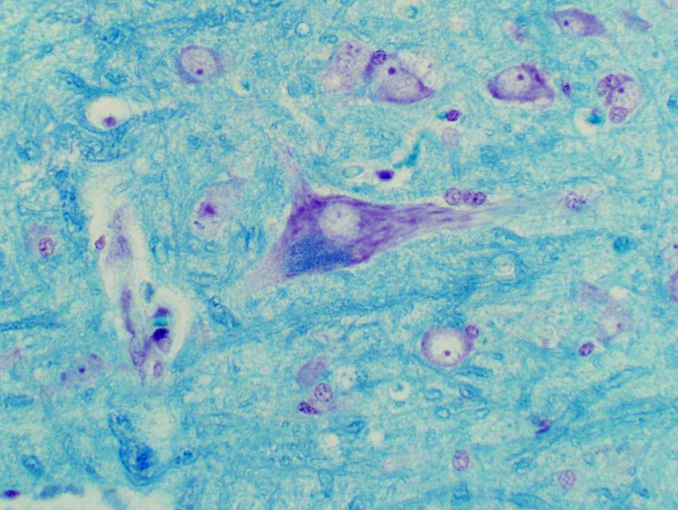
The Kluver-Barrera Stain is yet another combination method; it utilizes Luxol Fast Blue, Cresyl Violet, and a special component that's preferentially absorbed by the lipofuscin waste material that accumulates in neurons (see Exercise 3). In terms of delineating the morphology of neurons it isn't any better than an LFB and CV combination, but it has a very real value in being able to pinpoint the accumulations of lipofuscin.
Neurons are highly specialized and very long-lived cells that can't divide and have no capacity for endocytosis. The degraded and indigestible end products of turning over organelles is stored as granular material that stains a steel blue in a K-B preparation. In studies of neuronal degradation, this stain is exceptionally useful.
Canine Spinal Cord, 200x and 400x
| H&E | PAS | Masson's CT Stain | Verhoeff-van Gieson | Verhoeff-Masson | Mallory's CT Stain | Golgi Stain|
| Cresyl Violet | Cresyl Violet-Luxol Fast Blue | Kluver-Barrera | Fontana-Masson | Prussian Blue | Toluidine Blue|
|Osmium Tetroxide | Oil Red O | Sudan Black | Fluorescent & Enzymatic Tagging |