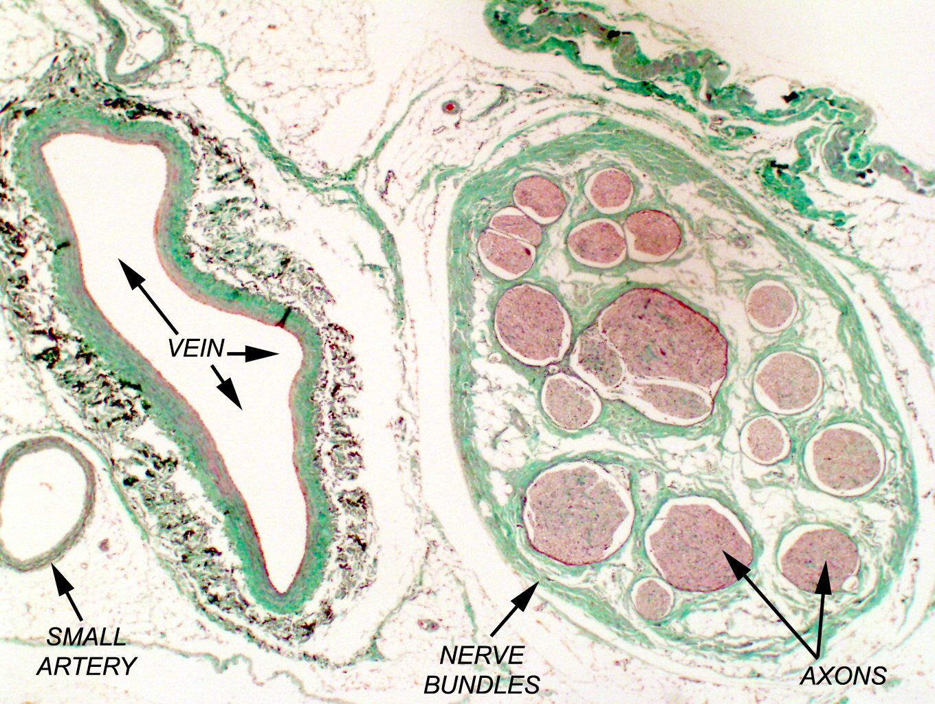
This is an example of Masson's Trichrome Stain for CT. There are many variations of this routine. In most of them collagenous components are stained a green or blue color and cytoplasm is varying shades of red. In this it's easy to make out the extensive collagenous fiber reinforcement of the wall of the large vein at left, and the CT wrappings of the bundles of nerve fibers at right. The higher magnification image shows the CT of the vein wall in detail.
Using this stain in combination with others for different CT components allows you to distinguish collagen from elastic fibers. The Masson stain is less often used than PAS, but it's immensely valuable in assessing such things as the extent of scarring and any other process that produces large amounts of collagen.
Femoral Artery & Femoral Nerve, Masson stain, paraffin section, 200x & 400x
| H&E | PAS | Masson's CT Stain | Verhoeff-van Gieson | Verhoeff-Masson | Mallory's CT Stain | Golgi Stain|
| Cresyl Violet | Cresyl Violet-Luxol Fast Blue | Kluver-Barrera | Fontana-Masson | Prussian Blue | Toluidine Blue|
|Osmium Tetroxide | Oil Red O | Sudan Black | Fluorescent & Enzymatic Tagging |
