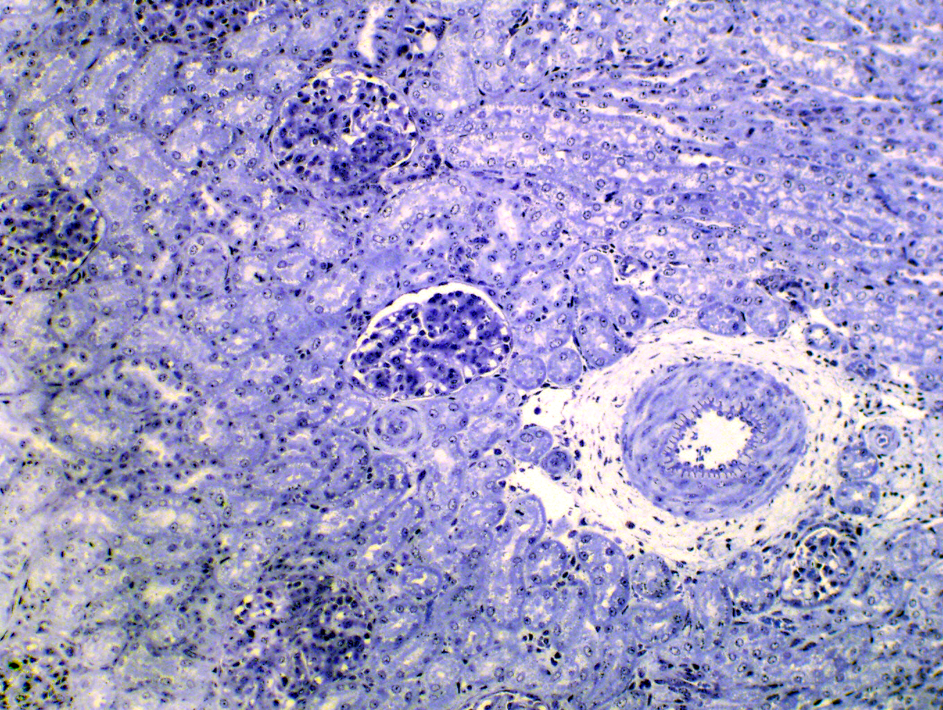

It's increasingly common to find sections stained with toluidine blue published in journals. TB is another basic dye. It's used as a quick stain for light microscopic "orientation sections" used by electron microscopists (EM images have so much magnification you have to look at sections in a light microscope first to figure out what it is you're going to be seeing in the EM). The pictures above are both of comparable areas of the kidney. The image at left was stained with TB: this process takes a few minutes as opposed to the hours needed for the H&E stain at right.
Because it's so often used nowadays, more and more journal articles have images made from TB-stained preparations, so it's important to be able to interpret these images as readily as the more common ones made with H&E. The TB stain shows general structure in the same way as H&E does, and you can directly compare these two sections, which are approximately at the same magnification, find many features in common. Like H&E, TB provides only minimal information about the chemical makeup of a tissue or organ.
TB is also used clinically. It has a strong affinity for the granules in mast cells, one of the wandering cells of connective tissue. TB is often requested as a specific stain for mast cell tumors.
Kidney, toluidine blue stain, 200x| H&E | PAS | Masson's CT Stain | Verhoeff-van Gieson | Verhoeff-Masson | Mallory's CT Stain | Golgi Stain|
| Cresyl Violet | Cresyl Violet-Luxol Fast Blue | Kluver-Barrera | Fontana-Masson | Prussian Blue | Toluidine Blue|
|Osmium Tetroxide | Oil Red O | Sudan Black | Fluorescent & Enzymatic Tagging |
